293 posters, 56 videos, 24 topics, 13 sessions, 658 authors, 215 institutions
ePostersLive by SciGen Technologies S.A. All rights reserved.
ISMICS 2025 Annual Meeting
28 - 31 May, 2025 | Istanbul, Turkey

D10
Simple Robotic Myxoma Resection Using An Ultrasonic Aspirator System
Poster Presenter
Authors
Part of Topic
Valve – Surgical Valves
Video
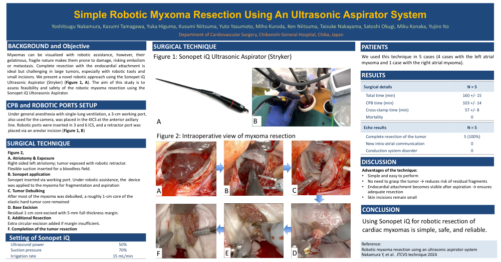
Performing myxoma resection through right mini- thoracotomy is reported to be less invasive and more efficient, with or without robot-assistance. Left atrial myxomas can be seen directly through right-side left atriotomy, but because myxomas are gelatinous and friable, they are easily damaged during surgical manipulation, potentially causing embolism or metastasis in the pulmonary veins or left ventricle. Therefore, resection of the tumor and its attachment to the endocardium as a single mass is recommended. However, this is difficult for large myxomas, especially when using robot assistance and small skin incisions. We report a novel technique in robot-assisted myxoma resection that uses the Sonopet iQ Ultrasonic Aspirator System (Stryker)
SURGICAL TECHNIQUE
Under general anesthesia using single-lung ventilation, a 3-cm working port also used for the robotic camera port was created in the fourth intercostal space on the left anterior axillary line. The left- and right-hand ports and robotic arms were placed in the third and sixth inter- costal spaces, respectively. A retractor port was placed through an areola incision. Cardiopulmonary bypass was established through the right femoral artery and femoral and jugular vein cannulation. Vacuum assistance was used for venous drainage (40-60 mm Hg). The ascending aorta was crossclamped using a Cygnet flexible clamp (Vitalitec Inc) with antegrade crystalloid cardioplegia delivery.
The myxoma was exposed with a robotic dynamic retractor through right side left atriotomy. A flexible sucker was inserted into the left atrium to achieve a bloodless field. A small sample of the myxoma was resected for immediate pathology confirmation. The patient- site surgeon inserted the Sonopet through the working port, positioning it against the myxoma with robotic assistance. The Sonopet’s outputs were set to 50% ultrasound power, 70% suction pressure, and 15 mL/minute irrigation rate. The myxoma was gradually fragmented and aspirated from the camera side. After most of the myxoma was debulked, a roughly 1-cm core of the elastic hard tumor core remained attached to the left atrial endocardium. The entire thickness of the left atrial endocardium at the myxoma attachment site was excised with a 5-mm margin. If there was concern about the margin size, an additional circular excision was made around the resected area.