293 posters, 56 videos, 24 topics, 13 sessions, 658 authors, 215 institutions
ePostersLive by SciGen Technologies S.A. All rights reserved.
ISMICS 2025 Annual Meeting
28 - 31 May, 2025 | Istanbul, Turkey

D35
Percutaneous Bicaval Balloon Occlusion In Minimally Invasive Right-sided Heart Surgery: Our Initial Experience
Poster Presenter
Authors
Beatrice Bacchi, Yasuhiko Kawaguchi, Ahmad Alli, Kendra Derry, Gianluigi Bisleri
Affiliations
Division of Cardiac Surgery, St. Michael’s Hospital, University of Toronto, Toronto, ON, Canada, Department of Anesthesiology & Pain Medicine, St. Michael's Hospital, University of Toronto, Toronto, ON, Canada
Part of Topic
Valve – Surgical Valves
PercutaneousBicaval Balloon Occlusion In Minimally Invasive Right-sided Heart Surgery: Our Initial Experience
Beatrice Bacchi1, Yasuhiko Kawaguchi1, Ahmad Alli2, Kendra Derry2, Gianluigi Bisleri1
1Division of Cardiac Surgery, St. Michael’s Hospital, University of Toronto, Toronto, ON, Canada
2Department of Anesthesiology & Pain Medicine, St. Michael's Hospital, University of Toronto, Toronto, ON, Canada
Objectives
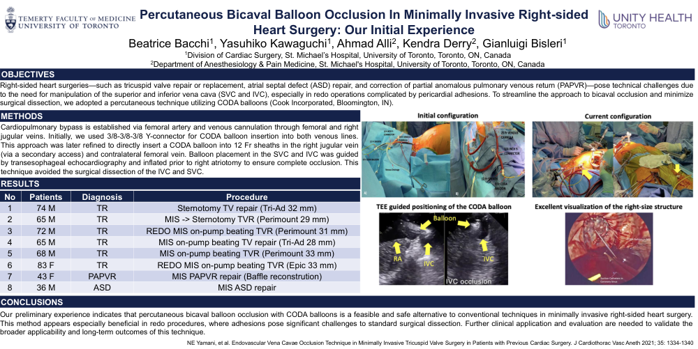
Right-sided heart surgeries—such as tricuspid valve repair or replacement, atrial septal defect (ASD) repair, and correction of partial anomalous pulmonary venous return (PAPVR)—pose technical challenges due to the need for manipulation of the superior and inferior vena cava (SVC and IVC), especially in redo operations complicated by pericardial adhesions. To streamline the approach to bicaval occlusion and minimize surgical dissection, we adopted a percutaneous technique utilizing CODA balloons (Cook Incorporated, Bloomington, IN).
Methods
Cardiopulmonary bypass is established via femoral artery and venous cannulation through femoral and right jugular veins. Initially, we used 3/8-3/8-3/8 Y-connector for CODA balloon insertion into both venous lines. This approach was later refined to directly insert a CODA balloon into 12 Fr sheaths in the right jugular vein (via a secondary access) and contralateral femoral vein. Balloon placement in the SVC and IVC was guided by transesophageal echocardiography and inflated prior to right atriotomy to ensure complete occlusion. This technique avoided the surgical dissection of the IVC and SVC.
Results
Table
Conclusions
Our preliminary experience indicates that percutaneous bicaval balloon occlusion with CODA balloons is a feasible and safe alternative to conventional techniques in minimally invasive right-sided heart surgery. This methodappears especially beneficial in redo procedures, where adhesions pose significantchallenges to standard surgical dissection. Further clinical application and evaluation are needed to validate the broader applicability and long-termoutcomes of this technique.