293 posters, 56 videos, 24 topics, 13 sessions, 658 authors, 215 institutions
ePostersLive by SciGen Technologies S.A. All rights reserved.
ISMICS 2025 Annual Meeting
28 - 31 May, 2025 | Istanbul, Turkey

D30
Totally Endoscopic Robotic Repair Of Unroofed Coronary Sinus Syndrome
Poster Presenter
Part of Topic
Valve – Surgical Valves
Video
Totally Endoscopic robotic repair of unroofed coronary sinus syndrome
Norihiko Ishikawa, Toru Koakutsu, Takafumi Horikawa, Daiki Yoshiyama, Shigeyuki Tomita, Toshiya Ohtsuka, Go Watanabe
Department of Cardiovascular Surgery, NewHeart Watanabe Institute, Tokyo JAPAN
Introduction
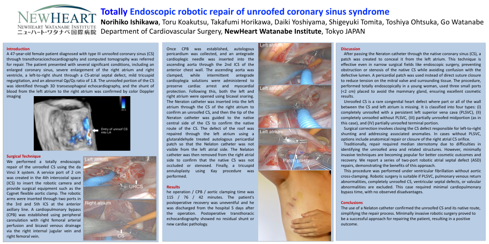
A 47-year-old female patient diagnosed with type III unroofed coronary sinus (CS) through transthoracicechocardiography and computed tomography was referred for repair. The patient presented with several significant conditions, including an enlarged coronary sinus, severe enlargement of the right atrium and right ventricle, a left-to-right shunt through a CS-atrial septal defect, mild tricuspid regurgitation, and an abnormal Qp/Qs ratio of 1.8. The unroofed portion of the CS was identified through 3D transesophageal echocardiography, and the shunt of blood from the left atrium to the right atrium was confirmed by color Doppler imaging
Surgical Technique
We performed a totally endoscopic repair of the unroofed CS using the da Vinci X system. A service port of 2 cm was created in the 4th intercostal space (ICS) to insert the robotic camera and provide surgical equipment such as the Cygnet flexible aortic clamp. The robotic arms were inserted through two ports in the 3rd and 5th ICS at the anterior axillary line. A cardiopulmonary bypass (CPB) was established using peripheral cannulation with right femoral arterial perfusion and bicaval venous drainage via the right internal jugular vein and right femoral vein.
Once CPB was established, autologous pericardium was collected, and an antegrade cardioplegic needle was inserted into the ascending aorta through the 2nd ICS of the anterior chest wall. The ascending aorta was clamped, while intermittent antegrade cardioplegia solutions were administered to preserve cardiac arrest and myocardial protection. Following this, both the left and right atrium were opened using bicaval snaring. The Neraton catheter was inserted into the left atrium through the CS of the right atrium to confirm an unroofed CS, and then the tip of the Neraton catheter was guided to the native central side of the CS to confirm the native route of the CS. The defect of the roof was repaired through the left atrium using a glutaraldehyde treated autologous pericardial patch so that the Nelaton catheter was not visible from the left atrial side. The Nelaton catheter was then removed from the right atrial side to confirm that the native CS was not occluded or stenosed. Finally, a tricuspid annuloplasty using Kay procedure was performed.
Results
he operation / CPB / aortic clamping time was 115 / 76 / 42 minutes. The patient's postoperative recovery was uneventful and he was discharged from the hospital 5 days after the operation. Postoperative transthoracic echocardiography showed no residual shunt or new cardiac pathology.
Discussion
After passing the Neraton catheter through the native coronary sinus (CS), a patch was created to conceal it from the left atrium. This technique is effective even in narrow surgical fields like endoscopic surgery, preventing obstruction or stenosis of the native CS while avoiding confusion with the defective lumen. A pericardial patch was used instead of direct suture closure to reduce tension on the mitral valve and surrounding tissue. The procedure, performed totally endoscopically in a young woman, used three small ports (<2 cm) placed to avoid the mammary gland, ensuring excellent cosmetic results.
Unroofed CS is a rare congenital heart defect where part or all of the wall between the CS and left atrium is missing. It is classified into four types: (I) completely unroofed with a persistent left superior vena cava (PLSVC), (II) completely unroofed without PLSVC, (III) partially unroofed midportion (as in this case), and (IV) partially unroofed terminal portion.
Surgical correction involves closing the CS defect responsible for left-to-right shunting and addressing associated anomalies. In cases without PLSVC, options include anatomical repair or closure of the right atrial CS orifice.
Traditionally, repair required median sternotomy due to difficulties in identifying the unroofed area and related structures. However, minimally invasive techniques are becoming popular for better cosmetic outcomes and recovery. We report a series of two-port robotic atrial septal defect (ASD) repairs, demonstrating the benefits of this approach.
This procedure was performed under ventricular fibrillation without aortic cross-clamping. Robotic surgery is suitable if PLSVC, pulmonary venous return abnormalities, completely unroofed CS, ventricular septal defects, or valvular abnormalities are excluded. This case required minimal cardiopulmonary bypass time, with no observed disadvantages.
Conclusions
The use of a Nelaton catheter confirmed the unroofed CS and its native route, simplifying the repair process. Minimally invasive robotic surgery proved to be a successful approach for repairing the patient, resulting in a positive outcome.