Loading...

293 posters, 56 videos, 24 topics, 13 sessions, 658 authors, 215 institutions

ePostersLive by SciGen Technologies S.A. All rights reserved.

ISMICS 2025 Annual Meeting

28 - 31 May, 2025 | Istanbul, Turkey

ISMICS 2025 Annual Meeting banner

D14

A Novel 'arrow Type' Mini-sternotomy: Comperative Study Of Biomechanical Stress Simulation With Conventional J And T Type Mini-sternotomy
Part of Topic

Aortic and Endovascular Therapies

A Novel 'Arrow Type' Mini-Sternotomy: Comparative Study Of Biomechanical Stress Simulation With Conventional J And T Type Mini-Sternotomy

Eyup Alper Saygili1, Murat Bastopcu2 , Gokhan Arslanhan2 , Zeynep Sila Ozcan3 , Anil Karaagac2 , Muharrem Kocyigit4 , Sahin Senay3 , Cem Alhan3

1 Acıbadem University School of Medicine, Istanbul, Turkey, 2 Acıbadem Maslak Hospital, Department of Cardiovascular Surgery, Istanbul, Turkey, 3 Acıbadem University School of Medicine, Department of Cardiovascular Surgery, Istanbul, Turkey, 4 Acıbadem University School of Medicine, Department of Anesthesiology, Istanbul, Turkey

Introduction

Sternotomy, particularly median sternotomy, is a cornerstone of cardiac surgery, offering excellent thoracic exposure and accounting for over a million procedures annually worldwide. However, it carries risks such as sternal wound infections, dehiscence, and prolonged recovery. To address these issues, minimally invasive approaches like partial sternotomies have gained popularity, providing quicker recovery and fewer complications.

The J-shaped sternotomy, widely used for aortic valve replacement and some coronary bypass surgeries, involves a midline incision extending laterally at the second or third intercostal space. This configuration balances adequate exposure with enhanced postoperative stability by preserving sternal integrity. Despite its advantages, the technique may limit access to posterior cardiac structures, prompting modifications to optimize exposure.

Here, we present a novel modification to the J sternotomy, incorporating a triangular extension at the distal end of the incision. This approach aims to enhance surgical access while preserving sternal stability and improving patient outcomes.

Surgical Technique

The modified Arrow Type J sternotomy procedure explanation is divided into steps:

1.Preparation: The surgical field is prepped and draped under sterile conditions, and the anatomical landmarks are identified. These include the suprasternal notch, the sternum's midline, the third and fourth intercostal spaces, xiphoid process, and 12th ribs.
●
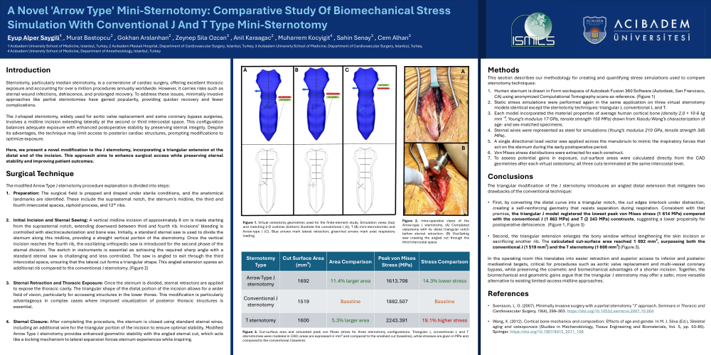
2.Initial Incision and Sternal Sawing: A vertical midline incision of approximately 8 cm is made starting from the suprasternal notch, extending downward between third and fourth rib. Incisions’ bleeding is controlled with electrocauterization and bone wax. Initially, a standard sternal saw is used to divide the sternum along this midline, providing a straight vertical portion of the sternotomy. Once the vertical incision reaches the fourth rib, the oscillating orthopedic saw is introduced for the second phase of the sternal division. The switch in instruments is essential as achieving the required sharp angle with a standard sternal saw is challenging and less controlled. The saw is angled to exit through the third intercostal space, ensuring that the lateral cut forms a triangular shape. This angled extension spares an additional rib compared to the conventional J sternotomy. (Figure 2)
●
3.Sternal Retraction and Thoracic Exposure: Once the sternum is divided, sternal retractors are applied to expose the thoracic cavity. The triangular shape of the distal portion of the incision allows for a wider field of vision, particularly for accessing structures in the lower thorax. This modification is particularly advantageous in complex cases where improved visualization of posterior thoracic structures is essential.
●
4.Sternal Closure: After completing the procedure, the sternum is closed using standard sternal wires, including an additional wire for the triangular portion of the incision to ensure optimal stability. Modified Arrow Type J sternotomy provides enhanced geometric stability with the angled sternal cut, which acts like a locking mechanism to lateral expansion forces sternum experiences while inspiring.
 

Methods

This section describes our methodology for creating and quantifying stress simulations used to compare sternotomy techniques:


1.Human sternum is drawn in Form workspace of Autodesk Fusion 360 Software (Autodesk, San Francisco, CA) using anonymized Computational Tomography scans as reference. (Figure 1)
2.Static stress simulations were performed again in the same application on three virtual sternotomy models identical except the sternotomy techniques: triangular J, conventional J, and T.
3.Each model incorporated the material properties of average human cortical bone (density 2.0 × 10⁻⁶ kg mm⁻³, Young’s modulus 17 GPa, tensile strength 150 MPa) drawn from Xiaodu Wang’s characterization of age- and sex-matched specimens.
4.Sternal wires were represented as steel for simulations (Young’s modulus 210 GPa, tensile strength 345 MPa).
5.A single directional load vector was applied across the manubrium to mimic the inspiratory forces that act on the sternum during the early postoperative period.
6.Von Mises stress distributions were extracted for each construct.
7.To assess potential gains in exposure, cut-surface areas were calculated directly from the CAD geometries after each virtual osteotomy; all three cuts terminated at the same intercostal level.


Conclusions

 
The triangular modification of the J sternotomy introduces an angled distal extension that mitigates two drawbacks of the conventional technique:


•First, by converting the distal curve into a triangular notch, the cut edges interlock under distraction, creating a self-reinforcing geometry that resists separation during respiration. Consistent with that premise, the triangular J model registered the lowest peak von Mises stress (1 614 MPa) compared with the conventional J (1 883 MPa) and T (2 243 MPa) constructs, suggesting a lower propensity for postoperative dehiscence.  (Figure 1, Figure 3)
•
•Second, the triangular extension enlarges the bony window without lengthening the skin incision or sacrificing another rib. The calculated cut-surface area reached 1 692 mm², surpassing both the conventional J (1 519 mm²) and the T sternotomy (1 600 mm²) (Figure 3).


In the operating room this translates into easier retraction and superior access to inferior and posterior mediastinal targets, critical for procedures such as aortic valve replacement and multi-vessel coronary bypass, while preserving the cosmetic and biomechanical advantages of a shorter incision. Together, the biomechanical and geometric gains argue that the triangular J sternotomy may offer a safer, more versatile alternative to existing limited-access midline approaches.


References

•Svensson, L. G. (2007). Minimally invasive surgery with a partial sternotomy “J” approach. Seminars in Thoracic and Cardiovascular Surgery, 19(4), 299–303. https://doi.org/10.1053/j.semtcvs.2007.10.004
•Wang, X. (2012). Cortical bone mechanics and composition: Effects of age and gender. In M. J. Silva (Ed.), Skeletal aging and osteoporosis (Studies in Mechanobiology, Tissue Engineering and Biomaterials, Vol. 5, pp. 53–85). Springer. https://doi.org/10.1007/8415_2011_108
ismics2025 banner
Copyright © 2015-2026, ePostersLive® SciGen is a registered trademark of SciGen Technologies S.A. Patent pending.

This website and third-party tools we use rely on cookies for the best user experience. By selecting "I agree", you agree to cookie usage as described in our Privacy Policy.