225 posters, 34 videos, 18 topics, 14 sessions, 251 authors, 55 institutions
ePostersLive by SciGen Technologies S.A. All rights reserved.
ISMICS 2024 Annual Meeting
29 May - 1 June, 2024 | Athens, Greece

D.138
Sternal Lifting Technique For Sternal Depression In Robotic Mitral Repair
Poster Presenter
Authors
Part of Topic
Valve - Surgical Valves
Sternal lifting technique for sternal depression in robotic mitral repair
Norihiko Ishikawa, Go Watanabe, Toru Koakutsu, Takafumi Horikawa, Ryuta Seguchi, Shigeyuki Tomita, Toshiya Ohtsuka
Department of Cardiovascular Surgery, NewHeart Watanabe Institute, Tokyo JAPAN
Introduction
Sternal depression, such as pectus excavatum (PE), is the most common congenital chest wall deformity. Although minimally invasive mitral valve surgery using a right intercostal approach has evolved in recent years, sternal depression makes these procedures difficult. We present our response to sternal depression in robotic mitral valve repair.
Materials and Surgical Technique
Patients who underwent robotic-assisted mitral repair using the da Vinci Surgical System (Intuitive surgical inc. Sunnyvale, CA) and sternal lifting technique at our institute from May 2014 to December 2022 were included. Patients with a sternum-to-vertebral distance (S/V distance) of less than 80 mm on preoperative CT scan routinely underwent sternal lift using the Electrical sternum lifting system (ESLS) at our institute.
Electrical sternum lifting system
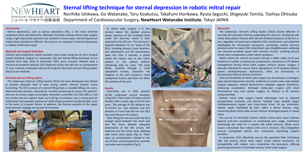
The Endoscopic Sternum Lifting System (ESLS) has been developed and utilized to provide adequate field of view during robotic internal thoracic artery harvesting. The ESLS consists of a sternal lifting hook, a movable lifting arm, and a table-mounted actuator, allowing for versatile positioning to access the patient's sternum at various angles and lengths. Remotely controlled, the ESLS offers a 150 mm stroke and can support loads up to 60 kg. Its actuator uses a screw-and-nut mechanism that provides mechanical self-locking to prevent accidental falls, even in the event of a power failure. In addition, the fulcrum position of the upper hook prevents slippage during sternal elevation.
In mitral valve surgery, a 3 mm incision above the xiphoid process allows insertion of the S-shaped hook under the sternum, which is then attached to the lift arm. The sternum is typically elevated 2-5 cm. Setup of the ESLS, including incision, hook insertion, arm attachment and lift optimization, takes less than 5 minutes. A photo shows the device next to the robot, docked to the patient without interfering with its arms. The hook blade, inserted through a small subxiphoid incision, maintains the integrity of the port position.a small subxiphoid incision and does not affect the position of the port.
Results
Sixty-three out of 1002 patients (6.3%) underwent sternal elevation using ESLS. There were 19 males and 44 females with a mean age of 50.9±14.0 years. The average of S/V distance was 72.4±8.9 cm. Two patients had S/V distance of more than 80 mm but ESLS was used because of scoliosis.
After lifting the sternum with the ESLS, enough space between the chest wall and the heart allowed adequate observation of the left atrium, left ventricle and the mitral valve, allowing safe mitral valve plasty (Fig 3). There were no complications related to the use of ESLS, and postoperative cosmetic outcomes were excellent (Fig 4).
Discussion
The Endoscopic Sternum Lifting System (ESLS) proved effective in securing the operative field by suspending the sternum, facilitating safe mitral valve plasty. Patients with sternal-vertebral distances of 80 mm or less, regardless of pulmonary embolism (PE) classification, were deemed challenging for intracardial maneuvers, prompting routine sternum elevation prior to repair. ESLS attachment was straightforward, widening the sternum-vertebrae space with minimal incision, enabling precise mitral valve manipulation under robotic guidance.
While criteria for sternal correction include a Haller index ≥3.25 and evidence of cardiac or pulmonary compression, consensus on PE patient management during mitral valve surgery remains elusive. Surgery is typically indicated for severe mitral regurgitation in PE patients without severe cardiopulmonary dysfunction, often via sternotomy or thoracotomy without sternal correction.
Technical feasibility of mitral valve surgery via sternotomy is contingent upon sternal depression severity; moderate to severe cases may impede surgical exposure. Lateral thoracotomy mitigates angular limitations, enhancing visualization. Although endoscopic surgery with small thoracotomy may rival robotic surgery, its efficacy in PE remains undocumented.
Robotic mitral valve surgery boasts advantages such as reduced perioperative mortality and shorter hospital stays despite longer cardiopulmonary bypass and cross-clamp times. At our institution, robotic repair, facilitated by ESLS, offers a lateral thoracic view, enhancing distal mitral valve access without prolonged bypass or clamp times.
Our pursuit of minimally invasive robotic mitral valve repair extends beyond port-only procedures to minimizing port usage, sometimes eliminating the need for a robotic left atrial retractor. While some repairs necessitate four robotic arms and a retractor, ESLS integration ensures unimpeded robotic arm movement, optimizing surgical precision.
In conclusion, ESLS effectively secures the operative field, facilitating safe and precise mitral valve repair under robotic assistance. Its compatibility with robotic arms streamlines the procedure, offering promising outcomes in minimally invasive mitral valve surgery.