225 posters, 34 videos, 18 topics, 14 sessions, 251 authors, 55 institutions
ePostersLive by SciGen Technologies S.A. All rights reserved.
ISMICS 2024 Annual Meeting
29 May - 1 June, 2024 | Athens, Greece

D.112
Endoscopic Transmitral Left Ventricular Pseudoaneurysm Repair
Poster Presenter
Authors
Part of Topic
Valve - Surgical Valves
Video
Endoscopic Transmitral Left Ventricular Pseudoaneurysm Repair
Mario Castillo-Sang, MD, Niem Ameer Khan, PA-C, Thomas Wilkinson, PA-C, John Creekmore, Prashant Nayak, MD.
St. Elizabeth Healthcare, Edgewood, KY, USA.
Abstract:
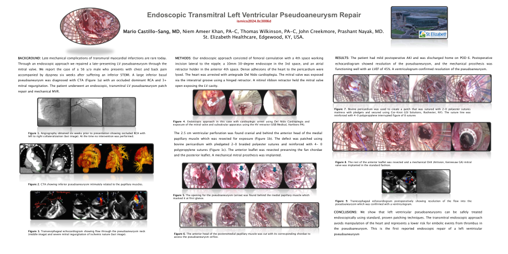
BACKGROUND: Late mechanical complications of transmural myocardial infarctions are rare today. Through an endoscopic approach we repaired a late-presenting LV pseudoaneurysm through the mitral valve. We report the case of a 56 y/o male who presents with chest and back pain accompanied by dyspnea six weeks after su"ering an inferior STEMI. A large inferior basal pseudoaneurysm was diagnosed with CTA (Figure 1a) with an occluded dominant RCA and 3+ mitral regurgitation. The patient underwent an endoscopic, transmitral LV pseudoaneurysm patch repair and mechanical MVR. METHODS: Our endoscopic approach consisted of femoral cannulation with a 4th space working incision lateral to the nipple; a 10mm 30-degree endoscope in the 3rd space, and an atrial retractor holder in the anterior 4th space. Dense adhesions of the heart to the pericardium were lysed. The heart was arrested with antegrade Del Nido cardioplegia. The mitral valve was exposed via the interatrial groove using a hinged retractor. A nitinol ribbon retractor held the mitral valve open exposing the LV cavity. The 2.5 cm ventricular perforation was found cranial and behind the anterior head of the medial papillary muscle which was resected for exposure (Figure 1b). The defect was patched using bovine pericardium with pledgeted 2-0 braided polyester sutures and reinforced with 4- 0 polypropylene sutures (Figure 1c). The anterior leaflet was resected preserving the fan chordae and the posterior leaflet. A mechanical mitral prosthesis was implanted. RESULTS: The patient had mild postoperative AKI and was discharged home on POD 6. Postoperative echocardiogram showed resolution of the pseudoaneurysm, and the mechanical prosthesis was functioning well with an LVEF of 45%. A ventriculogram confirmed resolution of the pseudoaneurysm. CONCLUSIONS: We show that left ventricular pseudoaneurysms can be safely treated endoscopically using standard, proven patching techniques. The transmitral endoscopic approach avoids manipulation of the heart and represents a lower risk for embolic events from thrombus in the pseudoaneurysm.
Figure 1. Angiography obtained six weeks prior to presentation showing occluded RCA with left to right collateralization (last image). At the time no intervention was performed.
Figure 2. CTA showing inferior pseudoaneurysm intimately related to the papillary muscles.
Figure 3. Transesophageal echocardiogram showing flowthrough the pseudoaneurysm neck (middle image) and severe mitral regurgitation of ischemic nature (last image).
Figure 4. Endoscopic approach in this case with cardioplegic arrest using Del Nido Cardioplegia and exposure of the mitral valve and subvalvular apparatus using the HV retractor (USB Medical, Hartboro PA).
Figure 5. The opening for the pseudoaneurysm (arrow) was found behind the medial papillary muscle which masked it at first glance.
Figure 6. The anterior head of the posteromedial papillary muscle was cut with its corresponding chordae to access the pseudoaneurysm orifice.
Figure 7. Bovine pericardium was used to create a patch that was sutured with 2-0 polyester sutures mattress with pledgets and secured using Cor-Knot (LSI Solutions, Rochester, NY). The suture line was reinforced with 4-0 polypropylene interrupted figure of 8 sutures
Figure 8. The rest of the anterior leaflet was resected and amechanical OnX (Artivion, Kennesaw GA) mitral valve was implanted in the standard fashion.
Figure 9. Tranesophageal echocardiogram postoperativelyshowing resolution of the flow into the pseudoaneurysm which was confirmed with a ventriculogram.