477 posters, 14 topics, 2,065 authors, 1,055 institutions
ePostersLive by SciGen Technologies S.A. All rights reserved.
The Joint EBJIS-MSIS 2026 Conference
17 - 19 September, 2026 | Porto, Portugal

EP015
Probe-Based Ultrasonication Removal of Staphylococcal Biofilm: In Vitro and Ex Vivo Proof of Concept
Poster Presenter
Authors
Bailey Fearing, Sarah Romereim, Aatif Jabbar, Matthew Shirley, Thomas Fehring, Jesse Otero
Affiliations
Musculoskeletal Institute, Atrium Health Wake Forest School of Medicine, Charlotte, United States, The Orthopedic Partners, Park City, United States, OrthoCarolina Hip and Knee Center, Charlotte, United States, Advocate Health
Part of Topic
Basic Science
Probe-Based Ultrasonication Removal of Staphylococcal Biofilm: In Vitro and Ex Vivo Proof of Concept
Introduction
Biofilm formation on implant surfaces is a major clinical challenge in periprosthetic joint infections
Ultrasonication has shown promise for disrupting biofilms, but efficacy on orthopaedic implant materials requires investigation
Presented here:
1. Quantification of ultrasonication removal of biofilm grown in vitro on orthopaedic materials
2. Qualitative observations of ex vivo biofilm removal using a novel mammalian model for staphylococcal knee PJI
Methods
The in vitro study:
Staphylococcus aureus cultured on four orthopaedic materials: bone cement (BC), polyethylene (PE), rough metal (RM, titanium alloy), and smooth metal (SM, polished cobalt chromium) (Fig. 1A-H)
Three ultrasonication protocols (pulse, 20-sec continuous, 60-sec continuous) with a submerged piezoelectric probe (Fig. 1I)
Biofilm presence/removal quantified by crystal violet absorbance assay
Colony forming units (CFU) remaining quantified by water bath sonication in culture media, serial dilution, agar plate culture, and colony counting
Statistics: Two-way ANOVAs with Tukey’s multiple comparisons and 95% confidence intervals were performed in GraphPad Prism 11.0.1.
Scanning electron microscopy (SEM) visualized biofilm changes
The in vivo study:
Novel rabbit model for knee PJI with a cementless 3D-printed porous titanium femoral unicondylar arthroplasty (Fig. 1J-L) [PMID: 42393525]
Intraarticular inoculation 2 weeks post-implant surgery with 2*105 CFU S. aureus
Biofilm developed in vivo for 2 weeks, distal end of the femur containing the implant removed (Fig. 1M) and placed in a sterile silicone mold (Fig. 1N), sterile PBS added, submerged piezoelectric probe ultrasonication performed, and SEM acquired
Negative control: no inoculation, no sonication
Positive control: inoculation, no sonication
Ex vivo biofilm removal: inoculation, 60 sec sonication
Results
Quantitative in vitro assays
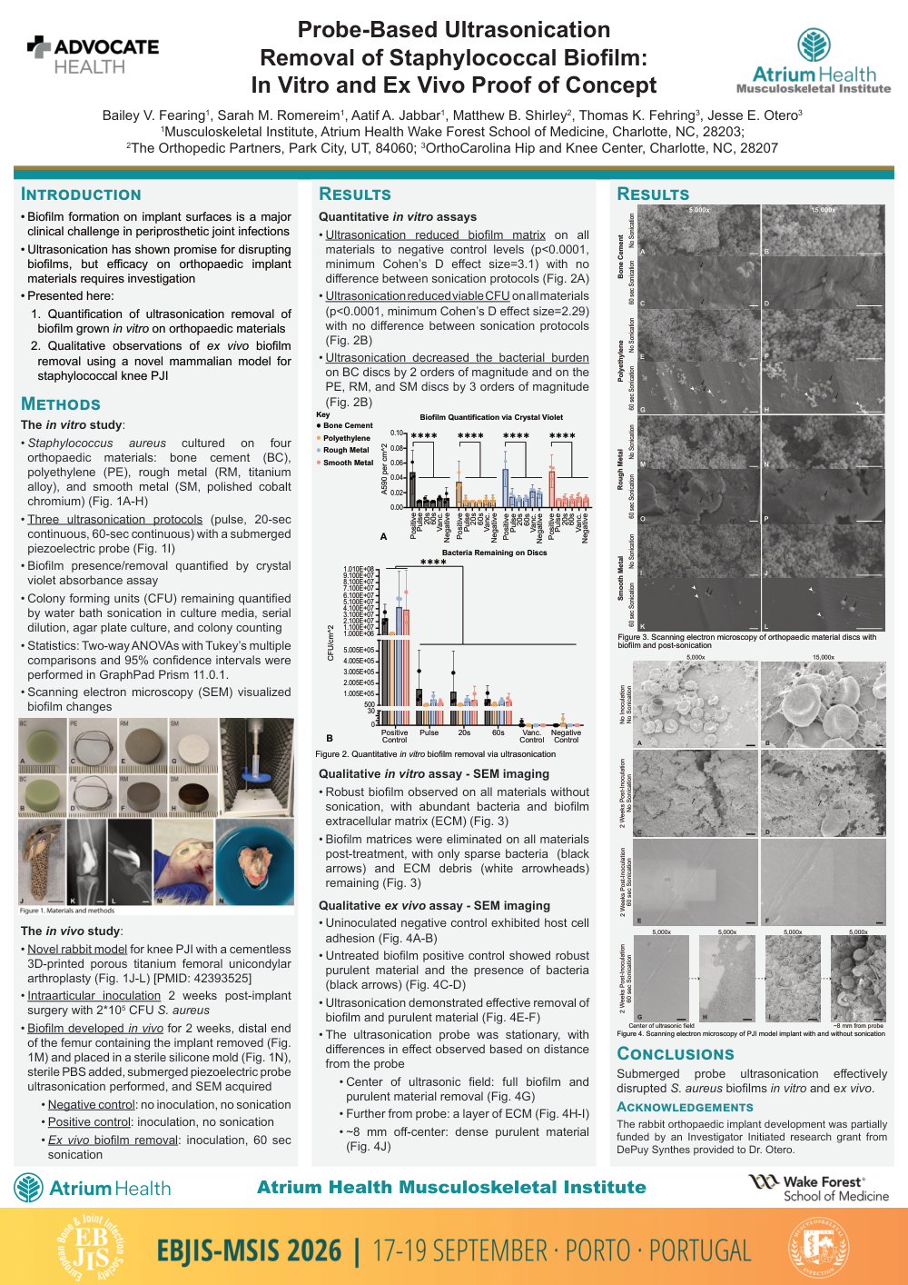
Ultrasonication reduced biofilm matrix on all materials to negative control levels (p<0.0001, minimum Cohen’s D effect size=3.1) with no difference between sonication protocols (Fig. 2A)
Ultrasonication reduced viable CFU on all materials (p<0.0001, minimum Cohen’s D effect size=2.29) with no difference between sonication protocols (Fig. 2B)
Ultrasonication decreased the bacterial burden on BC discs by 2 orders of magnitude and on the PE, RM, and SM discs by 3 orders of magnitude (Fig. 2B)
Qualitative in vitro assay - SEM imaging
Robust biofilm observed on all materials without sonication, with abundant bacteria and biofilm extracellular matrix (ECM) (Fig. 3)
Biofilm matrices were eliminated on all materials post-treatment, with only sparse bacteria (black arrows) and ECM debris (white arrowheads) remaining (Fig. 3)
Qualitative ex vivo assay - SEM imaging
Uninoculated negative control exhibited host cell adhesion (Fig. 4A-B)
Untreated biofilm positive control showed robust purulent material and the presence of bacteria (black arrows) (Fig. 4C-D)
Ultrasonication demonstrated effective removal of biofilm and purulent material (Fig. 4E-F)
The ultrasonication probe was stationary, with differences in effect observed based on distance from the probe
Center of ultrasonic field: full biofilm and purulent material removal (Fig. 4G)
Further from probe: a layer of ECM (Fig. 4H-I)
~8 mm off-center: dense purulent material (Fig. 4J)
Conclusions
Submerged probe ultrasonication effectively disrupted S. aureus biofilms in vitro and ex vivo.