92 posters, 1 audios, 567 authors, 81 institutions
ePostersLive by SciGen Technologies S.A. All rights reserved.
Data-driven Cancer Research Conference 2026
24-26 February 2026 | Edinburgh, Scotland

P81
Data-driven ex-vivo lung cancer detection using fibre-based fluorescence lifetime endomicroscopy
Poster Presenter
Authors
Affiliations
Part of Topic
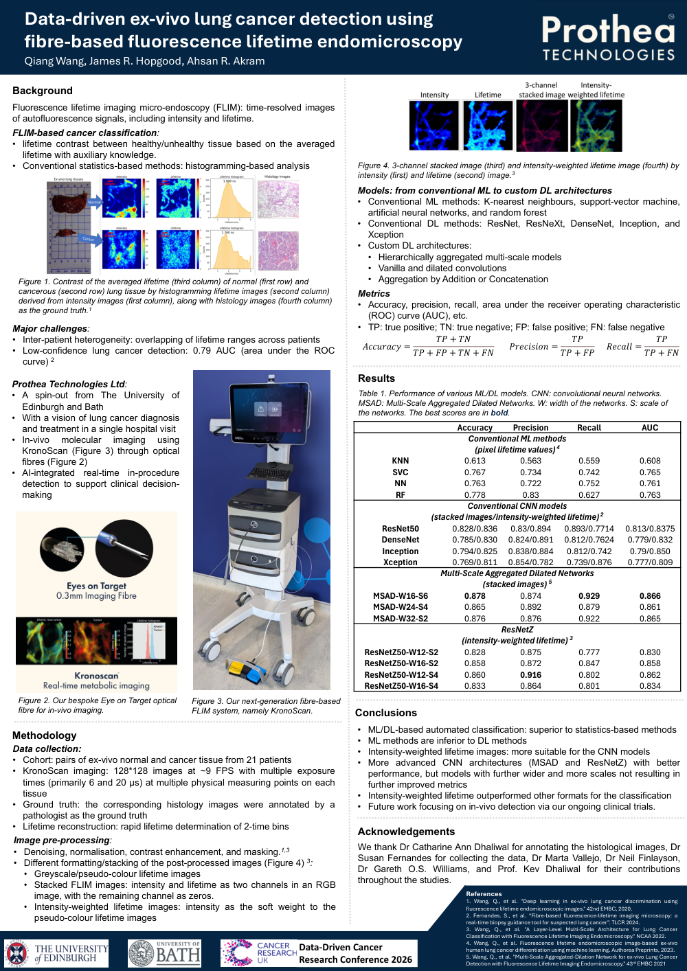
Early detection of lung cancer is critical for improving survival rates, yet current diagnostic workflows often rely on invasive biopsies and time-consuming histopathology. Fibre-based fluorescence lifetime endomicroscopy (FLIM) offers a minimally invasive, label-free imaging modality capable of probing tissue biochemical properties in real time, making it highly suitable for in-vivo applications during bronchoscopy. By measuring fluorescence decay dynamics, FLIM provides contrast sensitive to metabolic and structural changes associated with malignancy, enabling rapid tissue characterisation without exogenous dyes.
Over the past decade, a cutting-edge fibre-based FLIM system has been developed, enabling real-time imaging of small nodules in the distal lung. Using this system, we established a unique dataset comprising over 100,000 FLIM images from ex-vivo human lung tissue, representing a wide range of pathological conditions. Due to the complex nature of lifetime signals and tissue heterogeneity, conventional statistical-based analysis achieved only 0.79 area under the curve (AUC), highlighting the need for advanced computational approaches.
In this study, we present our efforts to improve classification performance using machine and deep learning (ML/DL) techniques. We first applied four conventional ML methods, namely K-nearest neighbour, support-vector classifier, neural network, and random forest, in parallel with principal component analysis. Using pixel lifetime values as input, these models achieved AUCs up to 0.765. We then improved the approach by introducing convolutional neural networks (CNNs), including ResNet and its variations, DenseNet, Inception, and Xception. By leveraging the optimal format for FLIM images, the AUC score increased to 0.858, demonstrating the advantage of CNNs in interpreting both spatial and temporal (lifetime) information. Finally, we optimised conventional CNN models by proposing multi-scale architectures and expanding the optimal image formats for FLIM data. This led to a further improvement of the AUC to 0.871. For all experiments, models were trained from scratch and tested on an independent cohort to ensure generalizability.
The integration of fibre-based FLIM with advanced deep learning architectures enables real-time, label-free identification of malignant lung tissue during bronchoscopy, eliminating the need for invasive biopsies and lengthy histopathology. By exploiting fluorescence lifetime as a sensitive biomarker of metabolic and structural alterations, combined with optimised multi-scale CNNs, clinicians can detect small distal nodules with high accuracy at the point of care. This capability reduces diagnostic delays, minimises unnecessary interventions, and supports immediate therapeutic decisions, ultimately improving patient outcomes. The fibre-based design ensures seamless compatibility with minimally invasive workflows, positioning this technology as a cornerstone for AI-driven precision diagnostics and accelerating its translation into routine clinical practice.
To further enhance diagnostic accuracy, future work will focus on integrating multi-modal information, such as patient demographics and radiological imaging, with FLIM data to exploit complementary features. Another critical advancement will be migrating the approach from ex-vivo analysis to real-time in-vivo deployment, enabling clinicians to make immediate, bedside decisions during bronchoscopy or surgical interventions.
Comments
Email is required but is not shown publicly.
Loading comments...