29 posters, 13 topics, 90 authors, 29 institutions
ePostersLive by SciGen Technologies S.A. All rights reserved.
Clinical Radiology Audit and Quality Improvement ePoster Competition 2025
18 NOVEMBER, 2025 | LONDON, UK

103
An Audit of Practice - Appropriate Vetting of CT Chest Requests in the Acute Setting
Poster Presenter
Authors
Affiliations
Part of Topic
Protocolling CT Chest in the acute setting – Are we getting it right?
P Nandra, E Chang, LNWUH NHS Trust
Introduction
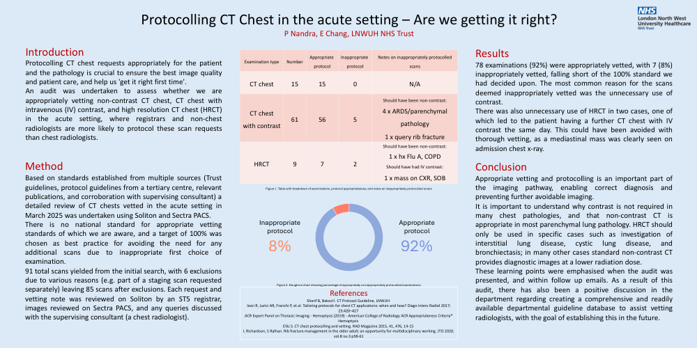
Protocolling CT chest requests appropriately for the patient and the pathology is crucial to ensure the best image quality and patient care, and help us 'get it right first time'. An audit was undertaken to assess whether we are appropriately vetting non-contrast CT chest, CT chest with intravenous (IV) contrast, and high resolution CT chest (HRCT) in the acute setting, where registrars and non-chest radiologists are more likely to protocol these scan requests than chest radiologists.
Method
Based on standards established from multiple sources (Trustguidelines, protocol guidelines from a tertiary centre, relevantpublications, and corroboration with supervising consultant) adetailed review of CT chests vetted in the acute setting inMarch 2025 was undertaken using Soliton and Sectra PACS.
There is no national standard for appropriate vettingstandards of which we are aware, and a target of 100% waschosen as best practice for avoiding the need for anyadditional scans due to inappropriate first choice ofexamination.
91 total scans yielded from the initial search, with 6 exclusionsdue to various reasons (e.g. part of a staging scan requestedseparately) leaving 85 scans after exclusions. Each request andvetting note was reviewed on Soliton by an ST5 registrar,images reviewed on Sectra PACS, and any queries discussedwith the supervising consultant (a chest radiologist).
Results
78 examinations (92%) were appropriately vetted, with 7 (8%)inappropriately vetted, falling short of the 100% standard wehad decided upon. The most common reason for the scansdeemed inappropriately vetted was the unnecessary use ofcontrast.
There was also unnecessary use of HRCT in two cases, one ofwhich led to the patient having a further CT chest with IVcontrast the same day. This could have been avoided withthorough vetting, as a mediastinal mass was clearly seen onadmission chest x-ray.
Conclusion
Appropriate vetting and protocolling is an important part of the imaging pathway, enabling correct diagnosis and preventing further avoidable imaging.
It is important to understand why contrast is not required in many chest pathologies, and that non-contrast CT is appropriate in most parenchymal lung pathology. HRCT should only be used in specific cases such as investigation of interstitial lung disease, cystic lung disease, and bronchiectasis; in many other cases standard non-contrast CT provides diagnostic images at a lower radiation dose.
These learning points were emphasised when the audit was presented, and within follow up emails. As a result of this audit, there has also been a positive discussion in the department regarding creating a comprehensive and readily available departmental guideline database to assist vetting radiologists, with the goal of establishing this in the future.