29 posters, 13 topics, 90 authors, 29 institutions
ePostersLive by SciGen Technologies S.A. All rights reserved.
Clinical Radiology Audit and Quality Improvement ePoster Competition 2025
18 NOVEMBER, 2025 | LONDON, UK
40
Evaluating CT aortogram requests and pre-test risk stratification in suspected acute aortic syndrome: a clinical audit
Poster Presenter
Authors
Affiliations
Part of Topic
CT and PET-CT
Evaluating CT aortogram requests and pre-test risk stratification in suspected acute aortic syndrome: a clinical audit
Mekha John, Helen Oliver, Mike Price
Introduction
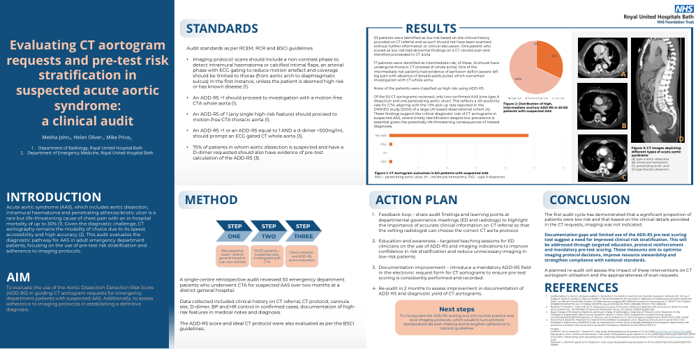
Acute aortic syndrome (AAS), which includes aortic dissection, intramural haematoma and penetrating atherosclerotic ulcer is a rare but life-threatening cause of chest pain with an in-hospital mortality of up to 30% (1). Given the diagnostic challenge, CT aortography remains the modality of choice due to its speed, accessibility and high accuracy (2). This audit evaluates the diagnostic pathway for AAS in adult emergency department patients, focusing on the use of pre-test risk stratification and adherence to imaging protocols.
Aim
To evaluate the use of the Aortic Dissection Detection Risk Score (ADD-RS) in guiding CT aortogram requests for emergency department patients with suspected AAS. Additionally, to assess adherence to imaging protocols in establishing a definitive diagnosis.
Standards
Audit standards as per RCEM3, RCR4 and BSCI5 guidelines
•An ADD-RS >1 or an ADD-RS equal to 1 AND a d-dimer >500ng/mL should prompt an ECG gated CTA.
A single-centre retrospective audit reviewed 50 emergency department patients who underwent CTA for suspected AAS over two months at a district general hospital.
Data collected included clinical history on CT referral, CT protocol, cannula site, D-dimer, BP and HR control in confirmed cases, documentation of high-risk features in medical notes and diagnosis.
The ADD-RS score and ideal CT protocol were also evaluated as per the BSCI guidelines.
Results
33 patients were identified as low risk based on the clinical history provided on CT referral and as such should not have been scanned without further information or clinical discussion. One patient who scored as low risk had abnormal findings on a CT carotid scan and therefore proceeded to CT aorta.
17 patients were identified as intermediate risk; of these, 16 should have undergone thoracic CT (instead of whole aorta). One of the intermediate risk patients had evidence of perfusion deficit (severe left leg pain with absence of dorsalis pedis pulse) which warranted investigation with CT whole aorta.
None of the patients were classified as high risk using ADD-RS.
Of the 50 CT aortograms reviewed, only two confirmed AAS (one type A dissection and one penetrating aortic ulcer). This reflects a 4% positivity rate for CTA, aligning with the <5% pick up rate reported in the DAShED study (2024) of a large UK-based observational cohort (3). These findings support the critical diagnostic role of CT aortograms in suspected AAS, where timely identification despite low prevalence is essential given the potentially life-threatening consequences of missed diagnoses.
Action Plan
The first audit cycle has demonstrated that a significant proportion of patients were low risk and that based on the clinical details provided in the CT requests, imaging was not indicated.
Documentation gaps and limited use of the ADD-RS pre-test scoring tool suggest a need for improved clinical risk stratification. This will be addressed through targeted education, protocol reinforcement and mandatory pre-test scoring. These measures aim to optimise imaging protocol decisions, improve resource stewardship and strengthen compliance with national standards.
A planned re-audit will assess the impact of these interventions on CT aortogram utilisation and the appropriateness of scan requests.