372 posters, 1 audios, 13 topics, 29 sessions, 1,035 authors, 449 institutions
ePostersLive by SciGen Technologies S.A. All rights reserved.
51st Annual Regional Anesthesiology and Acute Pain Medicine Meeting
April 16 - 18, 2026 | Phoenix, Arizona

2312232
Real-Time Ultrasound-Guided Spinal Anesthesia in a Patient with a Large Lumbar Subcutaneous Hemangioma
Poster Presenter
Part of Topic
Medically Challenging Cases
Real-Time Ultrasound-Guided Spinal Anesthesia in a Patient with a Large Lumbar Subcutaneous Hemangioma
Introduction
Spinal anesthesia is a common and effective technique for lower extremity procedures. However, anatomic abnormalities such as subcutaneous vascular lesions can complicate needle placement and increase bleeding risk. We report a case of successful real-time ultrasound-guided spinal anesthesia in a patient with a large lumbar subcutaneous hemangioma discovered immediately before elective total knee arthroplasty.
This case highlights the role of ultrasound guidance in safely navigating challenging spinal anatomy and avoiding vascular injury.
Materials and Methods
A 59-year-old male with hypertension, prediabetes, chronic kidney disease, and osteoarthritis presented for elective left total knee arthroplasty. Preoperative medical assessment did not
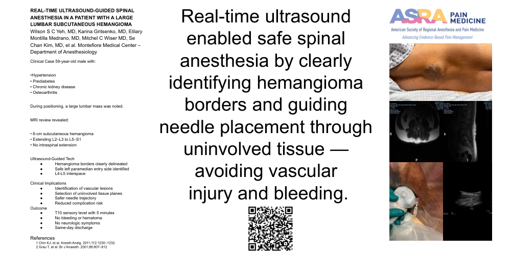
mention any spinal abnormalities. During positioning for spinal anesthesia, a large subcutaneous mass was noted over the lumbar region. After in depth chart review, an MRI image in the chart revealed a 6-cm subcutaneous hemangioma extending from L2–L3 to L5–S1 without intraspinal extension.
After discussing the risks and alternatives, informed consent was obtained for ultrasound-guided spinal anesthesia. Real-time ultrasound scanning (Sonosite xxx, curvilinear probe, 2–5 MHz) delineated the hemangioma and a safe left paramedian entry site. A 25G 5-inch Gertie Marx spinal needle was advanced under direct ultrasound visualization at the L4–L5 interspace until cerebrospinal fluid was obtained. As the case report contains no identifiable patient information, it was deemed exempt from IRB review under Montefiore Medical Center policy.
Results / Case Report
After sterile preparation and local infiltration, real-time ultrasound was used to guide the spinal needle trajectory, avoiding visible vascular channels. Clear visualization of the hemangioma borders allowed safe insertion through uninvolved tissue at L4–L5, with immediate cerebrospinal fluid return.
Hyperbaric 2,2 ml bupivacaine 0.5% with intrathecal 15 mcg of fentanyl was administered, achieving a T10 sensory block within 5 minutes. The surgery proceeded uneventfully, and no bleeding or hematoma was noted. The patient remained hemodynamically stable throughout the procedure and reported no neurological symptoms. He was discharged home the same day with full recovery. This case demonstrates that ultrasound can provide both diagnostic and procedural guidance in patients with unexpected subcutaneous vascular anomalies, reducing the risk of vascular puncture and enhancing procedural safety.
Discussion
Real-time ultrasound guidance can significantly improve safety in neuraxial anesthesia when vascular lesions are present. In this case, ultrasound enabled precise identification of a subcutaneous hemangioma and safe needle entry, avoiding potentially serious bleeding. As portable ultrasound technology advances, its use for spinal anesthesia should be considered in patients with altered anatomy or uncertain landmarks to minimize complications.
1. Chin KJ, et al. Ultrasound imaging facilitates spinal anesthesia in difficult cases. Anesth Analg. 2011;112(5):1230–1232.
2. Grau T, et al. Ultrasound imaging improves success and safety of spinal anesthesia in obstetrics. Br J Anaesth. 2001;86(5):807–812.