372 posters, 1 audios, 13 topics, 29 sessions, 1,035 authors, 449 institutions
ePostersLive by SciGen Technologies S.A. All rights reserved.
51st Annual Regional Anesthesiology and Acute Pain Medicine Meeting
April 16 - 18, 2026 | Phoenix, Arizona

2281003
Brachial Plexopathy After Clavicle Fracture Surgery
Poster Presenter
Authors
Affiliations
Part of Topic
Medically Challenging Cases
Brachial Plexopathy After Clavicle Fracture Surgery
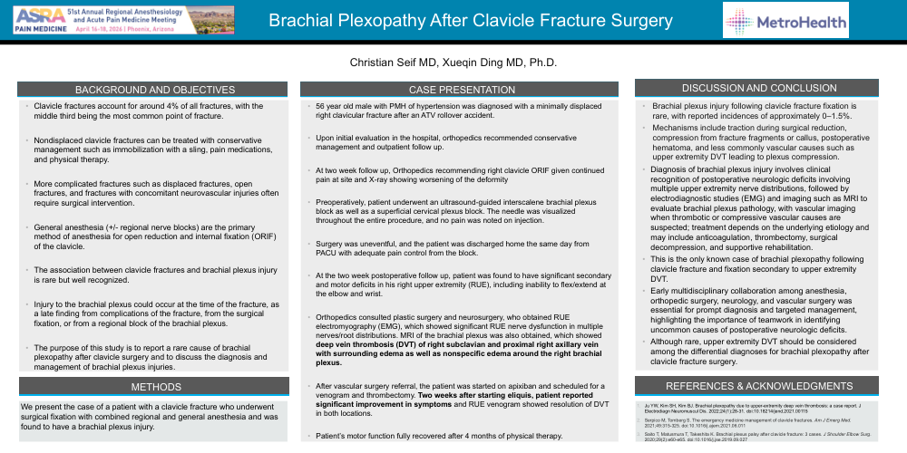
•Brachial plexus injury following clavicle fracture fixation is rare, with reported incidences of approximately 0–1.5%.
•Mechanisms include traction during surgical reduction, compression from fracture fragments or callus, postoperative hematoma, and less commonly vascular causes such as upper extremity DVT leading to plexus compression.
•Diagnosis of brachial plexus injury involves clinical recognition of postoperative neurologic deficits involving multiple upper extremity nerve distributions, followed by electrodiagnostic studies (EMG) and imaging such as MRI to evaluate brachial plexus pathology, with vascular imaging when thrombotic or compressive vascular causes are suspected; treatment depends on the underlying etiology and may include anticoagulation, thrombectomy, surgical decompression, and supportive rehabilitation.
•This is the only known case of brachial plexopathy following clavicle fracture and fixation secondary to upper extremity DVT.
•Early multidisciplinary collaboration among anesthesia, orthopedic surgery, neurology, and vascular surgery was essential for prompt diagnosis and targeted management, highlighting the importance of teamwork in identifying uncommon causes of postoperative neurologic deficits.
•Although rare, upper extremity DVT should be considered among the differential diagnoses for brachial plexopathy after clavicle fracture surgery.