383 posters, 2 videos, 1 audios, 8 topics, 28 sessions, 328 authors, 225 institutions
ePostersLive by SciGen Technologies S.A. All rights reserved.
50th Annual Regional Anesthesiology and Acute Pain Medicine Meeting
May 1 - 3, 2025 | Orlando, Florida

6493
UNUSUAL SOURCE OF SPINAL STENOSIS
Poster Presenter
Authors
Affiliations
Part of Topic
Medically Challenging Cases
Unusual source of spinal stenosis
Amelia Lerch, Jason McAloon, Diane Sierens, Gunar Subieta
Cook County Health
Introduction
Narrowing of the spinal canal or foramina is a common finding in spine imaging of the elderly and when symptoms of neurogenic claudication and/or cervical myelopathy are present the diagnosis of spinal stenosis is made. The etiologies are usually degenerative disc disease, ligamentum hypertrophy, a
combination of both and in rare cases an exogenous mass compression. We present an interesting case in which this mass effect was missed by the MRI and was finally diagnosed intraoperatively.
Materials and Methods
This presentation has no identifiable patient information; therefore, the case report is IRB exempt.
Patient informed consent was obtained to submit this case report.
Results/Case Report
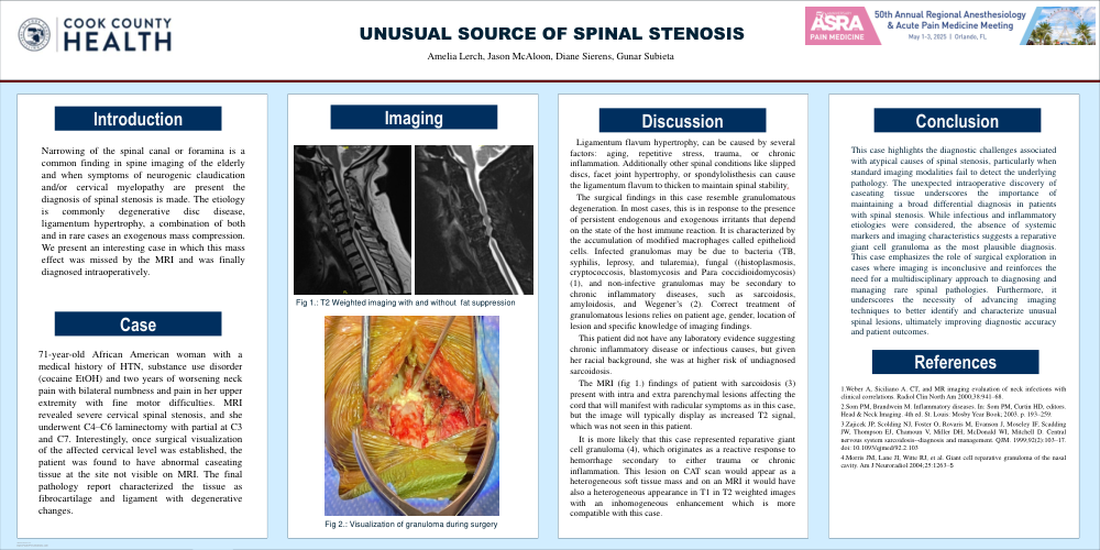
71-year-old African American woman with a medical history HTN, substance use disorder (cocaine EtOH) with two years of worsening neck pain with bilateral numbness and pain in her upper extremity with fine motor difficulties. MRI revealed severe cervical spinal stenosis, and she underwent C4–C6 laminectomy with partial at C3 and C7.
Interestingly, once surgical visualization of the affected cervical level was established, the patient was found to have abnormal caseating tissue at the site not visible on MRI. The final pathology report described the tissue as fibrocartilage and ligament with degenerative changes.
Discussion
Ligamentum flavum hypertrophy can be caused by a number of factors, including aging, repetitive stress, trauma or injury, chronic inflammation and other spinal conditions like slipped discs, facet joint hypertrophy, or spondylolisthesis can cause the ligamentum flavum to thicken to maintain spinal
stability.
The surgical findings in this case resembled granulomatous degeneration that in most cases occurs in response to the presence of persistent endogenous and exogenous irritants that depend on the state
of the host immune reaction.
Infected granulomas may be due to bacteria (TB, syphilis, leprosy and tularemia), fungal ((histoplasmosis, cryptococcosis, blastomycosis and para coccidioidomycosis) (1), and non-effective granulomas may be secondary to chronic inflammatory diseases, such as sarcoidosis, amyloidosis, and
Wegener’s (2).Correct treatment of granulomatous lesions relies on patient age, gender, location of lesion and specific knowledge of imaging findings.This patient did not have any laboratory evidence suggesting chronic inflammatory disease or infectious causes, but given her racial background, she was a higher risk of undiagnosed sarcoidosis. The MRI findings of patient with sarcoidosis (3) present with intra and extra parenchymal lesions affecting the cord that will manifest with radicular symptoms as in this case, but the image will display as increased T2 signal, which was not seen in this patient. It is more likely that this case represented reparative giant cell granuloma (4), which originates as a reactive response to hemorrhage secondary to either trauma or chronic inflammation. This lesion on CAT scan would appear as a heterogeneous soft tissue mass and on an MRI it would have also a heterogeneous appearance in T1 in T2 weighted images with a inhomogeneous enhancement which is more compatible with this case.
References
1. Weber A, Siciliano A. CT and MR imaging evaluation of neck infections with clinical correlations. Radiol Clin North Am 2000;38:941–68.
2. Som PM, Brandwein M. Inflammatory diseases. In: Som PM, Curtin HD, editors. Head & Neck Imaging. 4th ed. St. Louis: Mosby Year Book; 2003. p. 193–259.
3. Zajicek JP, Scolding NJ, Foster O, Rovaris M, Evanson J, Moseley IF, Scadding JW, Thompson EJ, Chamoun V, Miller DH, McDonald WI, Mitchell D. Central nervous system sarcoidosis--diagnosis and management. QJM. 1999;92(2):103–17. doi: 10.1093/qjmed/92.2.103
4. Morris JM, Lane JI, Witte RJ, et al. Giant cell reparative granuloma of the nasal cavity. Am J Neuroradiol 2004;25:1263–5