383 posters, 2 videos, 1 audios, 8 topics, 28 sessions, 328 authors, 225 institutions
ePostersLive by SciGen Technologies S.A. All rights reserved.
50th Annual Regional Anesthesiology and Acute Pain Medicine Meeting
May 1 - 3, 2025 | Orlando, Florida

6733
MRI EVIDENCE SHOWING EFFICACY OF AN EPIDURAL BLOOD-PATCH (EBP)
Poster Presenter
Part of Topic
Medically Challenging Cases
MRI Evidence Showing Efficacy of an Epidural Blood Patch
Introduction:
Cerebrospinal fluid (CSF) leakage can be imaged with Magnetic Resonance Imaging (MRI) techniques: the present case report provides direct evidence of CSF hypovolemia status post difficult spinal tap, successfully corrected with an epidural blood patch.
Patient Story:
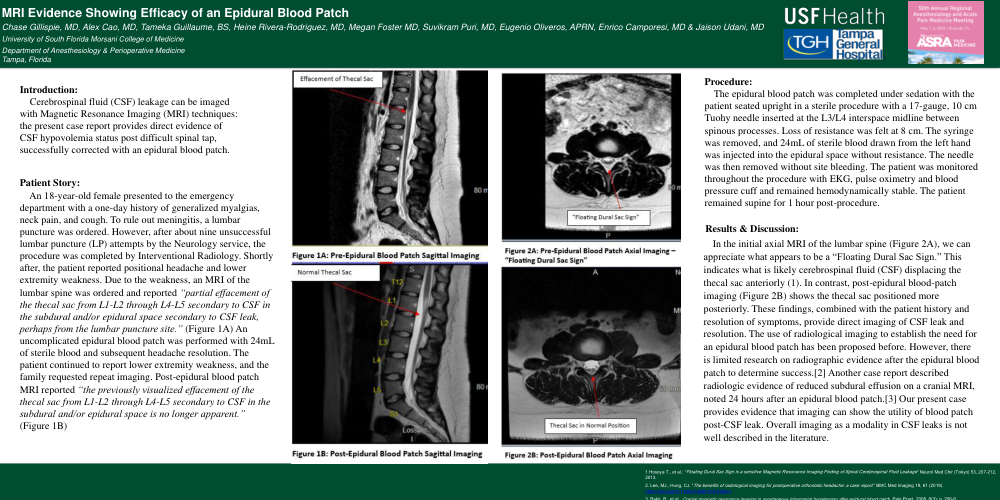
An 18-year-old female presented to the emergency department with a one-day history of generalized myalgias, neck pain, and cough. To rule out meningitis, a lumbar puncture was ordered. However, after about nine unsuccessful lumbar puncture (LP) attempts by the Neurology service, the procedure was completed by Interventional Radiology. Shortly after, the patient reported positional headache and lower extremity weakness. Due to the weakness, an MRI of the lumbar spine was ordered and reported “partial effacement of the thecal sac from L1-L2 through L4-L5 secondary to CSF in the subdural and/or epidural space secondary to CSF leak, perhaps from the lumbar puncture site.” (Figure 1A) An uncomplicated epidural blood patch was performed with 24mL of sterile blood and subsequent headache resolution. The patient continued to report lower extremity weakness, and the family requested repeat imaging. Post-epidural blood patch MRI reported “the previously visualized effacement of the thecal sac from L1-L2 through L4-L5 secondary to CSF in the subdural and/or epidural space is no longer apparent.” (Figure 1B)
Procedure:
The epidural blood patch was completed under sedation with the patient seated upright in a sterile procedure with a 17-gauge, 10 cm Tuohy needle inserted at the L3/L4 interspace midline between spinous processes. Loss of resistance was felt at 8 cm. The syringe was removed, and 24mL of sterile blood drawn from the left hand was injected into the epidural space without resistance. The needle was then removed without site bleeding. The patient was monitored throughout the procedure with EKG, pulse oximetry and blood pressure cuff and remained hemodynamically stable. The patient remained supine for 1 hour post-procedure.
Results & Discussion:
In the initial axial MRI of the lumbar spine (Figure 2A), we can appreciate what appears to be a “Floating Dural Sac Sign.” This indicates what is likely cerebrospinal fluid (CSF) displacing the thecal sac anteriorly (1). In contrast, post-epidural blood-patch imaging (Figure 2B) shows the thecal sac positioned more posteriorly. These findings, combined with the patient history and resolution of symptoms, provide direct imaging of CSF leak and resolution. The use of radiological imaging to establish the need for an epidural blood patch has been proposed before. However, there is limited research on radiographic evidence after the epidural blood patch to determine success.[2] Another case report described radiologic evidence of reduced subdural effusion on a cranial MRI, noted 24 hours after an epidural blood patch.[3] Our present case provides evidence that imaging can show the utility of blood patch post-CSF leak. Overall imaging as a modality in CSF leaks is not well described in the literature.