383 posters, 2 videos, 1 audios, 8 topics, 28 sessions, 328 authors, 225 institutions
ePostersLive by SciGen Technologies S.A. All rights reserved.
50th Annual Regional Anesthesiology and Acute Pain Medicine Meeting
May 1 - 3, 2025 | Orlando, Florida

6715
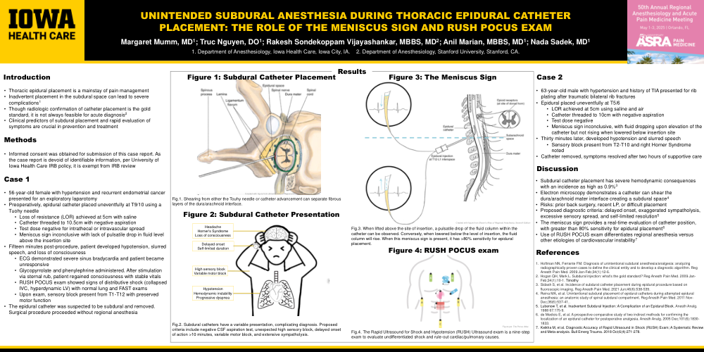
UNINTENDED SUBDURAL ANESTHESIA DURING THORACIC EPIDURAL CATHETER PLACEMENT: THE ROLE OF THE MENISCUS SIGN AND RUSH POCUS EXAM
Poster Presenter
Authors
Affiliations
Part of Topic
Medically Challenging Cases
UNINTENDED SUBDURAL ANESTHESIA DURING THORACIC EPIDURAL CATHETER PLACEMENT: THE ROLE OF THE MENISCUS SIGN AND RUSH POCUS EXAM
Margaret Mumm, MD1; Truc Nguyen, DO1; Rakesh Sondekoppam Vijayashankar, MBBS, MD2; Anil Marian, MBBS, MD1; Nada Sadek, MD1
Introduction:
•Thoracic epidural placement is a mainstay of pain management
•Inadvertent placement in the subdural space can lead to severe complications1
•Though radiologic confirmation of catheter placement is the gold standard, it is not always feasible for acute diagnosis2
•Clinical predictors of subdural placement and rapid evaluation of symptoms are crucial in prevention and treatment
Methods:
•Informed consent was obtained for submission of this case report. As the case report is devoid of identifiable information, per University of Iowa Health Care IRB policy, it is exempt from IRB review
Case1:
•56-year-old female with hypertension and recurrent endometrial cancer presented for an exploratory laparotomy
•Preoperatively, epidural catheter placed uneventfully at T9/10 using a Tuohy needle
•Loss of resistance (LOR) achieved at 5cm with saline
•Catheter threaded to 10.5cm with negative aspiration
•Test dose negative for intrathecal or intravascular spread
•Meniscus sign inconclusive with lack of pulsatile drop in fluid level above the insertion site
•Fifteen minutes post-procedure, patient developed hypotension, slurred speech, and loss of consciousness
•ECG demonstrated severe sinus bradycardia and patient became unresponsive
•Glycopyrrolate and phenylephrine administered. After stimulation via sternal rub, patient regained consciousness with stable vitals
•RUSH POCUS exam showed signs of distributive shock (collapsed IVC, hyperdynamic LV) with normal lung and FAST exams
•Upon exam, sensory block present from T1-T12 with preserved motor function
•The epidural catheter was suspected to be subdural and removed. Surgical procedure proceeded without regional anesthesia
Case2:
•63-year-old male with hypertension and history of TIA presented for rib plating after traumatic bilateral rib fractures
•Epidural placed uneventfully at T5/6
•LOR achieved at 5cm using saline and air
•Catheter threaded to 10cm with negative aspiration
•Test dose negative
•Meniscus sign inconclusive, with fluid dropping upon elevation of the catheter but not rising when lowered below insertion site
•Thirty minutes later, developed hypotension and slurred speech
•Sensory block present from T2-T10 and right Horner Syndrome noted
•Catheter removed, symptoms resolved after two hours of supportive care
Discussion:
•Subdural catheter placement has severe hemodynamic consequences with an incidence as high as 0.9%3
•Electron microscopy demonstrates a catheter can shear the dura/arachnoid mater interface creating a subdural space4
•Risks: prior back surgery, recent LP, or difficult placement
•Proposed diagnostic criteria: delayed onset, exaggerated sympatholysis, excessive sensory spread, and self-limited resolution5
•The meniscus sign provides a real-time evaluation of catheter position, with greater than 80% sensitivity for epidural placement6
•Use of RUSH POCUS exam differentiates regional anesthesia versus other etiologies of cardiovascular instability7