383 posters, 2 videos, 1 audios, 8 topics, 28 sessions, 328 authors, 225 institutions
ePostersLive by SciGen Technologies S.A. All rights reserved.
50th Annual Regional Anesthesiology and Acute Pain Medicine Meeting
May 1 - 3, 2025 | Orlando, Florida

6513
PNEUMOCEPHALUS SECONDARY TO AN EPIDURAL BLOCKADE: A CASE REPORT
Poster Presenter
Authors
Affiliations
Part of Topic
Medically Challenging Cases
PNEUMOCEPHALUS SECONDARY TO AN EPIDURAL BLOCKADE:
A CASE REPORT
BACKGROUND
Pneumocephalus: rare but serious complication of epidural anesthesia, potentially leading to severe neurological consequences.
It may present with acute headache, cranial nerve deficits, seizures, or altered mental status and may progress to brain herniation, brainstem compression, or persistent neurological deficits
METHODS
This case report describes a post-epidural pneumocephalus, with informed patient consent obtained and exemption from Institutional Review Board review per Hospital Israelita Albert Einstein policy.
RESULTS
Patient: 66-year-old female, ASA II
Surgery: Elective exploratory laparotomy for an ovarian mass
General anesthesia and Epidural blockade
Epidural block at T12-L1 + catheter secured at 13cm = test dose of 3mL of 1.5% lidocaine with epinephrine.
Intraoperatively: bolus of 5mL of 0.18% ropivacaine via catheter + continuous infusion of 2 mL.h-1 for 3 hours.
Patient was transfered to ICU and started on patient-controlled epidural analgesia (PCEA) with ropivacaine 0.2% and fentanyl 2μg/mL at 3mL/h. No intravenous PCA was used, and the patient did not activate optional bolus doses.
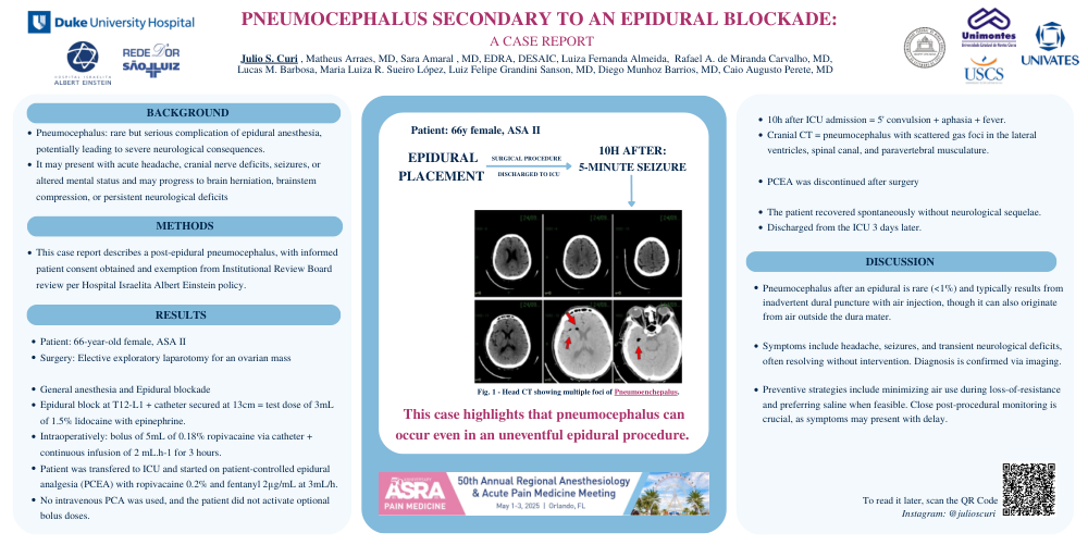
10h after ICU admission = 5' convulsion + aphasia + fever. Cranial CT = pneumocephalus with scattered gas foci in the lateral ventricles, spinal canal, and paravertebral musculature.
PCEA was discontinued after surgery
The patient recovered spontaneously without neurological sequelae. Discharged from the ICU 3 days later.
DISCUSSION
Pneumocephalus after an epidural is rare (<1%) and typically results from inadvertent dural puncture with air injection, though it can also originate from air outside the dura mater.
Symptoms include headache, seizures, and transient neurological deficits, often resolving without intervention. Diagnosis is confirmed via imaging.
Preventive strategies include minimizing air use during loss-of-resistance and preferring saline when feasible. Close post-procedural monitoring is crucial, as symptoms may present with delay.