383 posters, 2 videos, 1 audios, 8 topics, 28 sessions, 328 authors, 225 institutions
ePostersLive by SciGen Technologies S.A. All rights reserved.
50th Annual Regional Anesthesiology and Acute Pain Medicine Meeting
May 1 - 3, 2025 | Orlando, Florida

6702
PATCHING UP: MULTILEVEL, LARGE-VOLUME EPIDURAL BLOOD PATCH FOR INTRACTABLE HEADACHE FROM SPONTANEOUS INTRACRANIAL HYPOTENSION
Poster Presenter
Authors
Affiliations
Part of Topic
Medically Challenging Cases
Patching up: Multilevel, large-volume epidural blood patch for intractable headache from spontaneous intracranial hypotension
Joshua Brogan, MD1; Joss Thomas, MBBS, MPH, FCCP, MHA2; Clarence Shannon, MD2; Elena Averbach, MD2; Ratan K. Banik, MD, PhD2
1 Pain Medicine Fellowship, Department of Rehabilitation Medicine, University of Minnesota, Minneapolis, MN 2 Department of Anesthesiology, University of Minnesota, Minneapolis, MN
INTRODUCTION
Spontaneous intracranial hypotension (SIH) commonly results from cerebrospinal fluid (CSF) leaks, typically at the thoracic or cervicothoracic spine, with rare cases at the skull base
Causes include minor trauma or meningeal sac weakness, often linked to connective tissue disorders [1]
Hallmark symptom is an orthostatic headache [2][3]
When conservative treatments fail, epidural saline infusions or blood patches are recommended, for immediate volume replacement and sealing of dural defects [2]
Blood patch success rates for spontaneous CSF leaks are lower than for post-dural puncture headaches, often necessitating multiple treatments [4]
Evidence of the effectiveness of multilevel, large-volume (>20 mL) blood patches remains limited, however [5]
CASE REPORT
A 52-year-old male presented with 3 months of persistent orthostatic headache
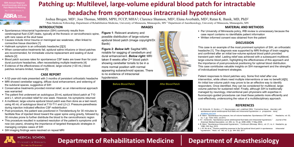
MRI showed cerebellar sagging, diffuse dural enhancement, and widening of the subdural spaces, suggestive of SIH
Conservative treatments provided minimal relief, so an interventional approach was warranted
The patient first underwent an autologous 20-mL epidural blood patch at T10 and L1, which provided relief for one week. However, his symptoms returned
A multilevel, large-volume epidural blood patch was then done as a last resort, using 40 mL of autologous blood at T10-T11 and L2-L3. Pressure-paresthesia during injection indicated effective CSF redistribution
Post-procedure, the patient was positioned in Trendelenburg for 30 minutes to facilitate flow of injected blood toward the upper spine using gravity, followed by 30 minutes prone to further distribute the blood to the cervicothoracic region
This procedure resulted in sustained resolution of the patient’s symptoms until now (six years), showing the importance of targeted therapeutic strategies in managing complex cases of SIH
SIH imaging findings were resolved on repeat MRI
IMAGES
Figure 1: Relevant anatomy and possible distribution of large-volume epidural blood patch (image copyright/Dr. Banik)
Figure 2: Below left: Sagittal MRI, notable for sagging of cerebellum and dural enhancement. Below right: MRI taken 8 weeks after 2nd blood patch showing cerebellar tonsils to be in a more normal position with normal appearing subarachnoid spaces. There is no evidence of intracranial hypotension.
MATERIAL AND METHODS
Per University of Minnesota policy, IRB review is unnecessary because the case report contains no identifiable patient information
Written informed consent was obtained from the patient
DISCUSSION
This case is an example of the most prominent symptom of SIH, an orthostatic headache [1]. The diagnosis was supported by MRI findings of brain sagging and confirmed after an initial low-volume epidural blood patch provided transient pain relief. Lasting relief was achieved with a subsequent multilevel, large-volume blood patch, highlighting the effectiveness of this approach and the importance of post-procedural positioning for optimal blood distribution. This case contributes valuable insights on SIH management and the strengths of individualized treatment strategies.
Patient responses to blood patches vary. Some find relief after one intervention, while others need multiple interventions or see no benefit [4][5]. An initial low-volume patch may prove to be an effective way to identify responders. Once identified, they can be considered for multilevel, large-volume patches for sustained relief. Finally, although SIH is traditionally managed by neurology, interventional pain physicians with expertise in fluoroscopic-guided procedures can treat these patients more efficiently and cost-effectively, underscoring the value of a multidisciplinary approach.