383 posters, 2 videos, 1 audios, 8 topics, 28 sessions, 328 authors, 225 institutions
ePostersLive by SciGen Technologies S.A. All rights reserved.
50th Annual Regional Anesthesiology and Acute Pain Medicine Meeting
May 1 - 3, 2025 | Orlando, Florida

6602
NOVEL USE OF A FENESTRATED NEEDLE FOR IPACK BLOCK
Poster Presenter
Authors
Affiliations
Part of Topic
Medically Challenging Cases
Title: Novel Use of a Fenestrated Needle for iPACK block
Authors: Amelia Updegraff, MD & Jacob Pollard, MD
Intro:
The iPACK block was first described in 2012 as a technique to control posterior knee pain in total knee arthroplasty1, which can be missed when using adductor canal blocks without peri-articular local anesthetic infiltration. Articular branches arise from the tibial and obturator nerves, travel between the popliteal artery and femur to innervate the posterior capsule2. By selectively targeting these terminal articular branches while sparing the tibial and common peroneal nerves, the iPACK can block sensation to the posterior knee while not affecting sensorimotor function of the lower leg, thus avoiding foot drop.
Materials & Methods:
As all patient identifying information has been removed, this is exempt from IRB approval per University of Utah’s policy. Patient was contacted via phone after being discharged and verbal consent was obtained to use this case for research and presentation purposes. Patient was informed that all identifying information would be omitted to protect privacy.
Case report:
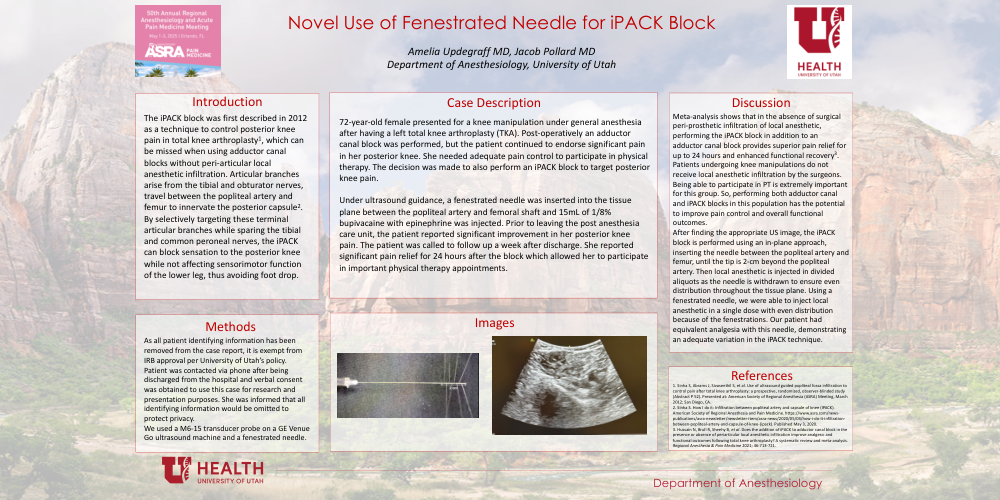
72-year-old female presented for a knee manipulation under general anesthesia after having a left total knee arthroplasty. Post-operatively an adductor canal block was performed, but the patient continued to endorse significant pain in her posterior knee. She needed adequate pain control to participate in physical therapy. The decision was made to also perform an iPACK block to target posterior knee pain. Under ultrasound guidance, a fenestrated needle was inserted into the tissue plane between the popliteal artery and femoral shaft and 15mL of 1/8% bupivacaine with epinephrine was injected. Prior to leaving the post anesthesia care unit, the patient reported significant improvement in her posterior knee pain. The patient was called to follow up a week after discharge. She reported significant pain relief for 24 hours after the block which allowed her to participate in important physical therapy appointments.
Discussion:
Meta-analysis of the iPACK block in the setting of total knee arthroplasty shows that in the absence of surgical peri-prosthetic infiltration of local anesthetic, performing the iPACK block in addition to an adductor canal block provides superior pain relief for up to 24 hours and enhanced functional recovery3. Patients undergoing knee manipulations do not receive local anesthetic infiltration by the surgeons and being able to participate in physical therapy is extremely important for this group. So, performing both adductor canal and iPACK blocks in this patient population has the potential to significantly improve not only their pain but also overall functional outcomes.
After finding the appropriate ultrasound image, the iPACK block is performed using an in-plane approach, inserting the needle in the plane between the popliteal artery and femur, until the tip is 2-cm beyond the popliteal artery. Once at this position, local anesthetic is injected in divided aliquots as the needle is withdrawn to ensure even distribution throughout the tissue plane. Using a fenestrated needle, we were able to inject local anesthetic in a single dose with even distribution because of the fenestrations at the end of the needle. Our patient had equivalent analgesia with this needle approach, demonstrating an adequate variation in the iPACK technique.