383 posters, 2 videos, 1 audios, 8 topics, 28 sessions, 328 authors, 225 institutions
ePostersLive by SciGen Technologies S.A. All rights reserved.
50th Annual Regional Anesthesiology and Acute Pain Medicine Meeting
May 1 - 3, 2025 | Orlando, Florida

6593
ERECTOR SPINAE CATHETER IN THE ICU FOR THORACOABDOMINAL AORTIC ANEURYSM REPAIR - A CASE STUDY
Poster Presenter
Part of Topic
Medically Challenging Cases
Erector Spinae Catheter in the ICU for Thoracoabdominal Aortic Aneurysm Repair – A Case Study
Erector spinae plane (ESP) blocks target a broad range of intercostal and spinal nerve rami in the paravertebral space, providing pain management after thoracic, vascular, abdominal and spine surgeries (1-2). Utilizing ESP blocks in high-risk patients, such as those undergoing thoracoabdominal aortic aneurysm (TAAA) repair, can be particularly useful. TAAA patients are frequently treated with anticoagulation, making other regional modalities, such as epidural or paravertebral anesthesia untenable (3, see table 1). ESP blocks offer a safer opioid-sparing alternative that can be performed even with anticoagulation. We present a case in which an ESP block in the cardiothoracic ICU (CTICU) effectively reduced opioid use and improved pain control following TAAA repair.
A 66-year-old woman with hypertension, type II diabetes mellitus and Loeys-Dietz Syndrome underwent a TAAA repair complicated by thrombosed renal artery stent in 2011. She recently represented with recurrent AAA distal to the repair, and underwent a thoracoabdominal aortic aneurysm plug with intercostal embolization. Postoperatively, she had significant pain, requiring a complex regimen of ketamine, acetaminophen, methocarbamol and oxycodone. She was re-admitted to the ICU for an endovascular leak and then underwent an open thoracoabdominal repair, left nephrectomy, left lower lobe wedge resection, and superior mesenteric artery, celiac artery, right renal artery bypass.
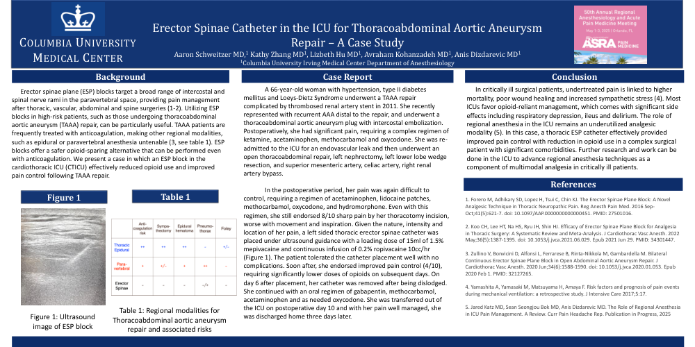
In the postoperative period, her pain was again difficult to control, requiring a regimen of acetaminophen, lidocaine patches, methocarbamol, oxycodone, and hydromorphone. Even with this regimen, she still endorsed 8/10 sharp pain by her thoracotomy incision, worse with movement and inspiration. Given the nature, intensity and location of her pain, a left sided thoracic erector spinae catheter was placed under ultrasound guidance with a loading dose of 15ml of 1.5% mepivacaine and continuous infusion of 0.2% ropivacaine 10cc/hr (Figure 1). The patient tolerated the catheter placement well with no complications. Soon after, she endorsed improved pain control (4/10), requiring significantly lower doses of opioids on subsequent days. On day 6 after placement, her catheter was removed after being dislodged. She continued with an oral regimen of gabapentin, methocarbamol, acetaminophen and as needed oxycodone. She was transferred out of the ICU on postoperative day 10 and with her pain well managed, she was discharged home three days later.
In critically ill surgical patients, undertreated pain is linked to higher mortality, poor wound healing and increased sympathetic stress (4). Most ICUs favor opioid-reliant management, which comes with significant side effects including respiratory depression, ileus and delirium. The role of regional anesthesia in the ICU remains an underutilized analgesic modality (5). In this case, a thoracic ESP catheter effectively provided improved pain control with reduction in opioid use in a complex surgical patient with significant comorbidities. Further research and work can be done in the ICU to advance regional anesthesia techniques as a component of multimodal analgesia in critically ill patients.