383 posters, 2 videos, 1 audios, 8 topics, 28 sessions, 328 authors, 225 institutions
ePostersLive by SciGen Technologies S.A. All rights reserved.
50th Annual Regional Anesthesiology and Acute Pain Medicine Meeting
May 1 - 3, 2025 | Orlando, Florida

6454
THORACIC EPIDURALS AND AN UNSUSPECTED COMPLICATION
Poster Presenter
Authors
Affiliations
Part of Topic
Medically Challenging Cases
RESULTS/CASE REPORT
66 year old male, with a BMI of 36 and a PMH of severe pulmonary hypertension (Group III), HTN, OSA, GERD, DMII, CAD s/p DES to the LAD who is S/p lung transplant and was intubated and sedated in the CVICU. The APS team was consulted to place a thoracic epidural POD1 for pain control but due to elevated INR (1.7) and thrombocytopenia (60k) secondary to cardio-pulmonary bypass the epidural was delayed until platelets reached above 100K and INR normalized which was on POD 4. The placement of the epidural was done at the level of the T7/T8 base of anatomical landmarks and the paramedian approach with loss of resistance to air was used with a Braun Perifix 18 G tuohy and 20 G catheter. The patient was in the sitting position with sedation held to get a good neurological exam, the placement was uneventful and with only one attempt. Once placed, the epidural infusion was set to run at 4 mL/hr with fentanyl 2mcg/mL and 1/8% bupivacaine. The patient's pain significantly improved, and he was able to be extubated the following day. The patient remained hemodynamically stable and had no motor weakness or sensory differences on physical exam.
The patient's pain was well controlled, and he was feeling well enough to work with PT/OT. On post catheter day 3 the patient walked with PT for the first time, when getting out of bed with PT the patient had a vasovagal event, became bradycardic and hypotensive. He was given multiple boluses of epinephrine 100mcg per mL and then proceeded to go into SVT which required a single cardioversion, and he was reintubated. The APS team was not notified of this event until the next day, and when the patient was examined, the epidural was still running and the patient remained hemodynamically stable. A physical exam was done with no noticeable weakness, but a sensory exam was difficult secondary to the patient being sedated and intubated. The catheter was aspirated, and clear fluid came back to fill the syringe, the fluid was tested for glucose which came back positive. The epidural was stopped immediately and eventually removed. The patient was extubated later that day and was discharged from the ICU the next day.
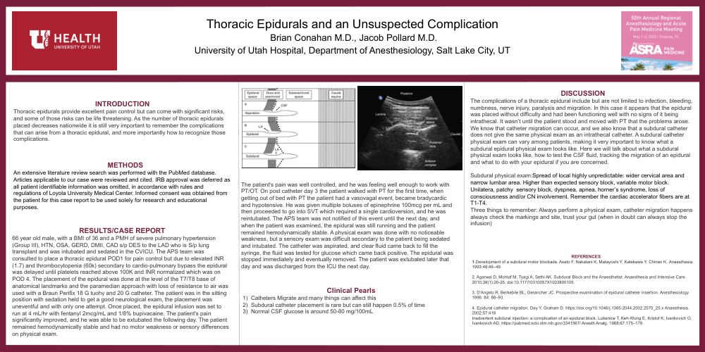
The complications of a thoracic ural include but are not limited to infection, bleeding, numbness, nerve injury, paralysis and migration. In this case it appears that the epidural was placed without difficulty and had been functioning well with no signs of it being intrathecal. It wasn’t until the patient stood and moved with PT that the problems arose. We know that catheter migration can occur, and we also know that a subdural catheter does not give the same physical exam as an intrathecal catheter. A subdural catheter physical exam can vary among patients, making it very important to know what a subdural epidural physical exam looks like. Here we will talk about what a subdural physical exam looks like, how to test the CSF fluid, tracking the migration of an epidural and what to do with your epidural if you are concerned.
Subdural physical exam:Spread of local highly unpredictable: wider cervical area and narrow lumbar area. Higher than expected sensory block, variable motor block. Unilatera, patchy sensory block, dyspnea, apnea, horner’s syndrome, loss of consciousness and/or CN involvement. Remember the cardiac accelerator fibers are at T1-T4.
Three things to remember: Always perform a physical exam, catheter migration happens always check the markings and site, trust your gut (when in doubt can always stop the infusion)