383 posters, 2 videos, 1 audios, 8 topics, 28 sessions, 328 authors, 225 institutions
ePostersLive by SciGen Technologies S.A. All rights reserved.
50th Annual Regional Anesthesiology and Acute Pain Medicine Meeting
May 1 - 3, 2025 | Orlando, Florida

6794
UNINTENDED MEDIAL CORD BLOCK FOLLOWING PECS I AND II NERVE BLOCKS
Poster Presenter
Part of Topic
Medically Challenging Cases
Unintended Medial Cord Block Following PECS I and II Nerve Blocks: A Case
Report Highlighting Anatomical Considerations and Technique Optimization
Alejandro Hallo, MD1; Adarsh Menon, MD1; Alexander Pekurovsky, MD2;
1 Anesthesiology Department - Rutgers New Jersey Medical School; 2 Cooperman Barnabas Medical Center, Livingston, New Jersey
Introduction
• PECS I and II nerve blocks target the pectoral nerves and
thoracic intercostal nerves, providing anesthesia to the
pectoralis major and minor muscles as well as adjacent
thoracic wall structures.
• Despite their superficial approach and clearly defined
ultrasound landmarks, complications can arise from the
unintended spread of local anesthetic to nearby structures.
• This report presents a rare case of medial cord block
following a PECS I and II procedure.
Case 1
References
• A 35-year-old female underwent an augmentation mammoplasty
under general anesthesia with endotracheal intubation without
intraoperative complications. Her prior surgical and medical
histories were unremarkable.
• Post operatively, she received an ultrasound guided bilateral PECS I
and II nerve block
• Block technique and medication:
• 15 ml of local anesthetic solution comprised of 20 ml Liposomal
Bupivacaine (Exparel ) + 20 ml of bupivacaine 0.25% + 20 ml of
normal saline, was injected between the pectoralis major and
minor muscles.
• The PECS II block involved an additional 15 ml of the local
anesthetic solution noted above to be injected between the
pectoralis minor and serratus anterior muscles.
Several hours after block administration, the patient reported
paresthesia in the right arm. Physical examination revealed motor
weakness and sensory deficits areas innervated by the medial cord of
the brachial plexus.
• There was a degree of patchiness with regards to symptoms
distribution, though overall symptoms were localized to the C8 and T1
dermatomes but were not strictly defined by of the median, ulnar,
median cutaneous nerve distributions.
• The patient was discharged home with telephone follow-ups. She was
advised to elevate the upper extremity. The nerve block gradually
wore off over several days and fully resolved by day five.
Conclusion
• This report illustrates a rare complication of medial cord block
following PECS I and II procedures.
• While these techniques are generally regarded as safe and effective,
their anatomical proximity to the brachial plexus highlights the
necessity for precise technique and judicious volume management.
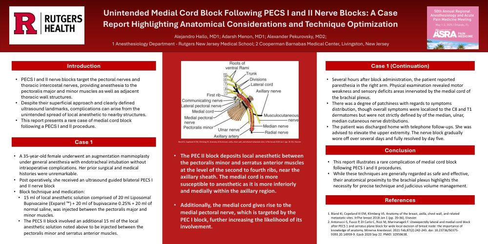
The PEC II block deposits local anesthetic between
the pectoralis minor and serratus anterior muscles
at the level of the second to fourth ribs, near the
axillary sheath. The medial cord is more
susceptible to anesthetic as it is more inferiorly
and medially within the axillary region.
• Additionally, the medial cord gives rise to the
medial pectoral nerve, which is targeted by the
PEC I block, further increasing the likelihood of its
involvement.