383 posters, 2 videos, 1 audios, 8 topics, 28 sessions, 328 authors, 225 institutions
ePostersLive by SciGen Technologies S.A. All rights reserved.
50th Annual Regional Anesthesiology and Acute Pain Medicine Meeting
May 1 - 3, 2025 | Orlando, Florida

6678
INTRAOPERATIVE DIAGNOSIS OF ACUTE DVT WHILE PERFORMING ADDUCTOR CANAL BLOCK
Poster Presenter
Authors
Affiliations
Part of Topic
Medically Challenging Cases
INTRAOPERATIVE DIAGNOSIS OF ACUTE DVT WHILE PERFORMING ADDUCTOR CANAL BLOCK
INTRODUCTION
Venous duplex doppler is the first-line method of identifying deep vein thrombosis (DVT); however, bedside compression ultrasonography offers similar accuracy when scanning patients with suspected DVT1,2. Performing lower extremity nerve blocks provides a convenient opportunity to also quickly screen for DVT in patients with concerning risk factors or symptoms.
We present a patient scheduled for total knee arthroplasty (TKA) who was found to have acute DVT on ultrasound scanning of the femoral vein while preparing for an adductor canal block.
CASE PRESENTATION
In May 2024, a 72-year-old male with a history of factor V Leiden mutation complicated by DVT and pulmonary embolism (PE) following gallbladder surgery 20 years prior, treated with six months of anticoagulation, presented for elective TKA. Other history included hypercholesterolemia, gastroesophageal reflux disease, and obstructive sleep apnea.
On the day of surgery, his vitals, physical exam, labs, and echocardiogram were normal. A stress test showed 1 mm ST depressions without inducible ischemia, and CTA revealed minimal obstructive disease. The anesthetic plan included adductor canal saphenous, anterior femoral cutaneous, genicular, and infiltration between the popliteal artery and capsule of the knee (IPACK) nerve blocks followed by spinal anesthesia and arterial line placement.
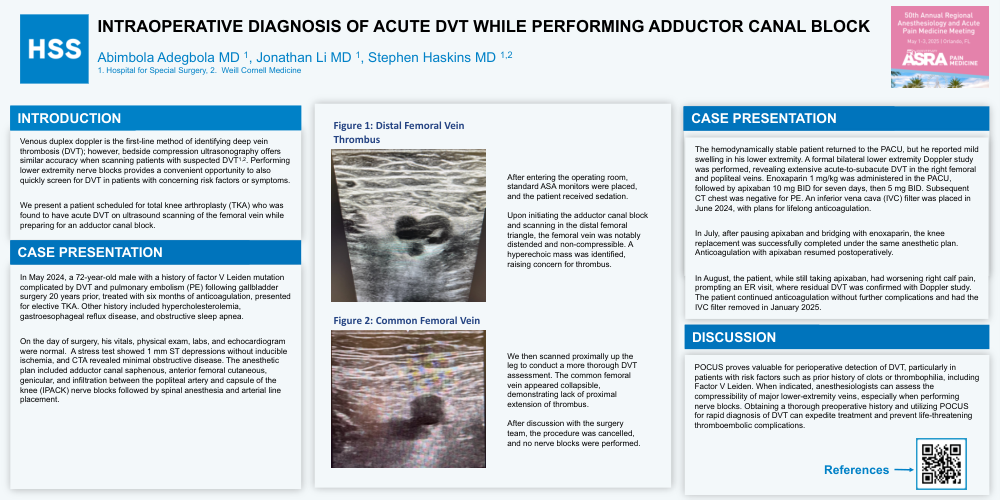
After entering the operating room, standard ASA monitors were placed, and the patient received sedation.
Upon initiating the adductor canal block and scanning in the distal femoral triangle, the femoral vein was notably distended and non-compressible. A hyperechoic mass was identified, raising concern for thrombus.
We then scanned proximally up the leg to conduct a more thorough DVT assessment. The common femoral vein appeared collapsible, demonstrating lack of proximal extension of thrombus.
After discussion with the surgery team, the procedure was cancelled, and no nerve blocks were performed
The hemodynamically stable patient returned to the PACU, but he reported mild swelling in his lower extremity. A formal bilateral lower extremity Doppler study was performed, revealing extensive acute-to-subacute DVT in the right femoral and popliteal veins. Enoxaparin 1 mg/kg was administered in the PACU, followed by apixaban 10 mg BID for seven days, then 5 mg BID. Subsequent CT chest was negative for PE. An inferior vena cava (IVC) filter was placed in June 2024, with plans for lifelong anticoagulation.
In July, after pausing apixaban and bridging with enoxaparin, the knee replacement was successfully completed under the same anesthetic plan. Anticoagulation with apixaban resumed postoperatively.
In August, the patient, while still taking apixaban, had worsening right calf pain, prompting an ER visit, where residual DVT was confirmed with Doppler study. The patient continued anticoagulation without further complications and had the IVC filter removed in January 2025.
DISCUSSION
POCUS proves valuable for perioperative detection of DVT, particularly in patients with risk factors such as prior history of clots or thrombophilia, including Factor V Leiden. When indicated, anesthesiologists can assess the compressibility of major lower-extremity veins, especially when performing nerve blocks. Obtaining a thorough preoperative history and utilizing POCUS for rapid diagnosis of DVT can expedite treatment and prevent life-threatening thromboembolic complications.