457 posters, 2 videos, 2 audios, 8 topics, 45 sessions, 401 authors, 265 institutions
ePostersLive by SciGen Technologies S.A. All rights reserved.
49th Annual Regional Anesthesiology and Acute Pain Medicine Meeting
March 21-23, 2024 | The Hilton Bayfront San Diego | San Diego, California

5476
ERECTOR SPINAE PLANE BLOCK WITH SUPERFICIAL CERVICAL PLEXUS BLOCK FOR ANALGESIA IN A PATIENT UNDERGOING FIRST RIB RESECTION
Poster Presenter
Authors
Affiliations
Part of Topic
Medically Challenging Cases
Title - Erector spinae plane block with superficial cervical plexus block for analgesia in a patient undergoing first rib resection
Authors - Clayton F. Kolle, MD (1), Dustin Smith, MD (2), Justin Swengel, MD (3)
Introduction - Thoracic outlet syndrome (TOS)is a group of disorders in which there is compression of or damage to the neurovascular structures at the thoracic outlet, the space between the clavicle and the first rib. The mainstay of surgical management for all types of TOS is first rib resection. Moderate to severe postoperative pain is often anticipated for this surgical procedure. While opioids are often used for analgesia, their well-described side effects have led physicianstosearchfor alternative analgesicmodalities such as regional analgesicprocedures to help with postoperative pain control. At our institution, the supraclavicular approach is used most often for first rib resections. For our case, the surgical team requested that the brachial plexus was not blocked so they could monitor motor function and sensation postoperatively.Thus, the decision was made by the anesthesia team to perform a superficial cervical plexus block (SCPB) plus an erector spinae plane block (ESPB) to help manage postoperative pain.
Case Report - A 35-year-old male ASA grade II with a new diagnosis of neurogenic thoracic outlet syndrome presented for a first rib resection. The patient had a past medical history of general anxiety disorder, and a remote history of substance abuse. Of note, due to the patient’s history of substance abuse, he requested to avoid all opioid medications if possible, during the perioperative period.
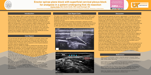
Informed consent for a SCPB and an ESPB was obtained from the patient.After the appropriate ASA monitors were placed, the patient got into a sitting position for the ESPB. The left mid-thoracic area was prepped,and ultrasound was used to identify the traverse process at T2 using a linear probe.A 22g 80 mm insulated needle was inserted in-plane, and incremental injection of 22 ml's local anesthetic solution containing0.5% Ropivacaine with Decadron 4 mg was seen spreading in the inter fasciae plane between the transverse process and erectorspinaemuscles. The patient was then laid supine for the SCPB, and the left supraclavicular area was prepped.With the head and neck turned away from the operative site, the suprascapular neck area was scanned with ultrasound to reveal neural elements of SCP. A 22g 50 mm insulated needle was inserted in-plane and incremental injection of 8 ml’s of the same solution was seen spreading around the neural elements.
The patient was induced with intravenouslidocaine, fentanyl, propofol, and succinylcholine followed by orotracheal intubation.At surgical incision, no change in the patient’s hemodynamic markers was observed. Anesthesia was maintained with sevoflurane. Intraoperatively, the patient received ketorolac,dexmedetomidine, andondansetron.
The surgery was completed uneventfully. The patient was followed up in PACU where he reported a pain score of 8/10 in his left scapula and down his left arm. The patient denied pain at his incisional site. After a one hour stay in PACU the patient was transferred to the floor.
On the floor the patient received scheduled acetaminophen, gabapentin, ketorolac, methocarbamol, and lidocaine patches. Patient consistently reported pain scores of 8/10 down his arm during his hospitalization, but the pain was never unbearable enough to use PRN opioid medications. The patient was then discharged after two days.
Discussion - In thesupraclavicular approach, the incision is approximately 1-2 cm above the clavicle, followed by the dissection of the anterior and middle scalene muscles and the resection of the first rib.This approach affects an anatomical region abundant in sensory nerves, including high cervical spinal nerves via the cervical plexus, cervicothoracic spinal nerves to the brachial plexus, and high thoracic spinal nerves. These networks of nervessupply innervation to the scalene and subclavian muscles, the clavicle, as well as innervation of the ribs and intercostal spaces. Considering the high volume of sensory nerves in this area, selecting the right analgesic technique becomes crucial when planning a regional nerve block.There is limited literature on regional analgesic methods specific to this approach.
The ESPB was chosendue to the relationship of the three superficial layers of the peri spinous musculature and the dorsal rami contributions from the spine. The mechanism of this block is not fully understood, but it is attributed to the possible spread to the paravertebral space and laterally to the intercostal nerves. It has been used for analgesia in surgeries involving chest wall and axilla. Our assumption was that this block would not eliminate post operative pain but would help to reduce it and minimize opioid use. Additionally,the SCPB was chosen to provide analgesia to the incision site in the peri-clavicular and neck region. This block is most often associated with analgesia for carotid endarterectomy surgery. However, its utility has grown into other procedures such asclavicleand submandibular surgeries, as well as neck dissections.
In our case report, the ESPB and SCPBseemed to at least contribute to the required level of pain control throughout his hospitalization period. Although he still had somepain mostly down his arm, associated with irritation of the brachial plexus, the nerve blocks enhanced the patient's comfortand made it possible to remain opioid-free during the perioperative period.However, the true efficacyand potential of the ESP block in reducing opioid requirements can only be ascertained through future prospective comparative studies.
References:
Dengler NF, Pedro MT, Kretschmer T, Heinen C, Rosahl SK, Antoniadis G. Neurogenic Thoracic Outlet Syndrome. DtschArztebl Int. 2022 Oct 28;119(43):735-742. doi: 10.3238/arztebl.m2022.0296. PMID: 35978467; PMCID: PMC9975980.
Suzuki T, Kimura H, Matsumura N, Iwamoto T. Surgical Approaches for Thoracic Outlet Syndrome: A Review of the Literature. J Hand Surg Glob Online. 2022 May 5;5(4):577-584. doi: 10.1016/j.jhsg.2022.04.007. PMID: 37521542; PMCID: PMC10382894.
Barros M, Carvalho T, Pires AC, Teixeira G, Cardoso H. EffectivePostoperativePain Management in ThoracicOutletSyndromeSurgery: The Role oftheErectorSpinae Plane Block. Cureus. 2023 Nov 17;15(11):e48944. doi: 10.7759/cureus.48944. PMID: 38106791; PMCID: PMC10725572.
Nagdev, Arun. “Superficial cervical plexusblock”. Highland EM Ultrasound, CC BY-NC-ND 4.0, 2012, http://highlandultrasound.com/superficial-cervical-plexus-block.