457 posters, 2 videos, 2 audios, 8 topics, 45 sessions, 401 authors, 265 institutions
ePostersLive by SciGen Technologies S.A. All rights reserved.
49th Annual Regional Anesthesiology and Acute Pain Medicine Meeting
March 21-23, 2024 | The Hilton Bayfront San Diego | San Diego, California

5284
SURGICAL PLACEMENT AND POST-OP MANAGEMENT OF LUMBAR MULTIFIDUS AND ERECTOR SPINAE NERVE BLOCK CATHETERS FOR COMPLEX LUMBAR SURGERY
Poster Presenter
Authors
Affiliations
Part of Topic
Medically Challenging Cases
SURGICAL PLACEMENT AND POST-OP MANAGEMENT OF LUMBAR MULTIFIDUS AND ERECTOR SPINAE NERVE BLOCK CATHETERS FOR COMPLEX LUMBAR SURGERY
John N Yi M.D. 1
Chase Foster M.D. 2, Eric Heinz M.D. 1,
Michael Rosner M.D. 2, Moshe Chinn M.D.1
INTRO
•Significant postoperative pain from complex lumbar spinal fusions originate from trauma around facet joint/pedicle, alongside muscle dissection leading to multifidus/erector spinae muscle spasm.
•This case study showcases unique collaboration between Anesthesia, Chronic Pain, and Neurosurgery to figure out a “continuous/adjustable medical branch block” in acute postoperative period.
•Due to wide field of surgery, ESPB theoretically could be placed under direct visualization.
•
MATERIALS & METHODS
•Patient informed consent obtained.
•ESPB Catheter Placement Technique
•Proceduralist scrubbed into surgical field for performance under strict sterile conditions due to open incision.
•Cobb elevator was used to retract erector spinae/multifidus muscle laterally off base of transverse process.
•4-inch BBraun 18g block needle was placed through skin above incision (cranial-to-caudal trajectory) until touching Cobb.
•Block needle slid down cobb until resting securely on transverse process.
•Cobb removed, and catheter advanced along the transverse process.
•Final visualization was made to ensure good placement of nerve block catheter. Catheters were capped with sterile alligator clamp.
•After surgical closure, catheter were bolused with local anesthetic (bupivacaine).
RESULTS/CASE REPORT
•73 y/o F with hx of chronic back pain & lumbar stenosis with prior L5-S1 fusion presenting for L5/S1 MAST TLIF.
•Prior surgery complicated by severe opioid-induced constipation requiring further hospitalization. Opioid-sparing pain management requested by patient.
•Intraoperative general anesthesia maintained with TIVA (prop/ketamine/precedex). Intraoperative ESPB placed under ultrasound for this 2nd surgery, and Ropi 0.25%, 20cc bolused.
•Catheters connected to Sapphire pumps. Postoperative course otherwise uncomplicated and catheters bolused/pulled on POD2.
•Patient presented again 2.5 months later with further revision surgery, this time full L2-S1 decompression. Due to large open incision, decision made this time to place L4 catheters under direct visualization. Bolused bupi 0.25%, 10cc.
•Patient discharged home on POD4 with good pain control and not requiring IV push PRN opioids since surgery.
DISCUSSION
•Patient described ESPB technique as “only thing that really worked”, with objective pain scores reflective of that statement.
•Little/no PRN opioid medications were needed to control pain.
•Titration of ESPB catheters worth was worth focusing, as larger doses led to significant and bothersome leg numbness/heaviness.
•Limitation of this novel method is that it requires technical expertise and high degree of trust/collaboration with surgeons.
•Since this case, technique has undergone slight modification and has been performed for other patients.
•Of note, the technique was taught to our neurosurgical colleagues as an option for postoperative pain management for future complex lumbar spine surgery.
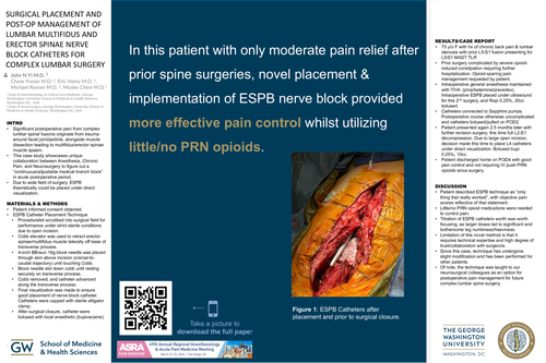
In this patient with only moderate pain relief after prior spine surgeries, novel placement & implementation of ESPB nerve block provided more effective pain control whilst utilizing little/no PRN opioids.