457 posters, 2 videos, 2 audios, 8 topics, 45 sessions, 401 authors, 265 institutions
ePostersLive by SciGen Technologies S.A. All rights reserved.
49th Annual Regional Anesthesiology and Acute Pain Medicine Meeting
March 21-23, 2024 | The Hilton Bayfront San Diego | San Diego, California

5207
INTRAOPERATIVE USE OF ULTRASOUND FOR PERIPHERAL NERVE LOCALIZATION FOR DECOMPRESSION
Poster Presenter
Authors
Affiliations
Part of Topic
Medically Challenging Cases
Intraoperative Use of Ultrasound for Peripheral Nerve Localization For Decompression
Patient Case: A 72 year old woman with a history of hip replacement with an anterior approach presented to the pain clinic with complaints of left sided lateral thigh pain for 3 years. She was found to have a positive left sided Tinel’s at the LFCN just inferior to the ASIS. A diagnosis of meralgia paresthetica was made. A left lateral femoral cutaneous nerve (LFCN) block provided 100% relief for four hours. Due to continued pain despite medical therapy, decompression of the left LFCN was scheduled with neurosurgery.
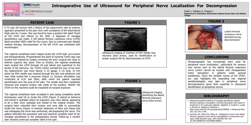
The regional anesthesia team helped locate the LFCN both pre-incision and intraoperatively. Preoperatively the point of maximal Tinel’s sign was located and marked by surgery overlying the prior surgical site close to anterior superior iliac spine. Prior to incision, the regional anesthesia team located the LFCN through US just lateral and superficial to the border of the Sartorius. Her Tinel’s (which overlaid the scar of her prior hip replacement) was more lateral. A 22 gauge, 5 cm long, 10 mm active tip RFA needle was inserted through the skin and advanced until near what looked like a neuroma (Figure 1). Sensory stimulation was conducted at 0.2V and 50Hz, after which the patient reported paresthesias over the area of her pain. The needle was then completely removed and surgical incision was made at the location. Neither the LFCN nor the neuroma could be visualized on surgical exposure.
The regional anesthesia team scrubbed in and (using completely sterile techniques) used US to locate the LFCN (Figure 2) lateral to Sartorius and traced it cephalad where its trajectory was more lateral, appearing to lie a little more cephalad and medial to the original incision. The surgical team extended their incision and were able to successfully locate the nerve (Figure 3) wherein dissection of thick scar tissue that was entrapping the nerve was performed, decompressing the nerve. The patient tolerated the procedure well and reported complete relief of her meralgia paresthesia in the postoperative period. Follow-up 2 months later showed continued complete relief of her pain.