457 posters, 2 videos, 2 audios, 8 topics, 45 sessions, 401 authors, 265 institutions
ePostersLive by SciGen Technologies S.A. All rights reserved.
49th Annual Regional Anesthesiology and Acute Pain Medicine Meeting
March 21-23, 2024 | The Hilton Bayfront San Diego | San Diego, California

5591
LUNG ULTRASOUND FOR EARLY DETECTION OF PNEUMOTHORAX, UNCOMMON CONDITION FOLLOWING SHOULDER SURGERY AND INTERSCALENE NERVE BLOCK
Poster Presenter
Authors
Affiliations
Part of Topic
Safety/QA/QI Projects
Introduction
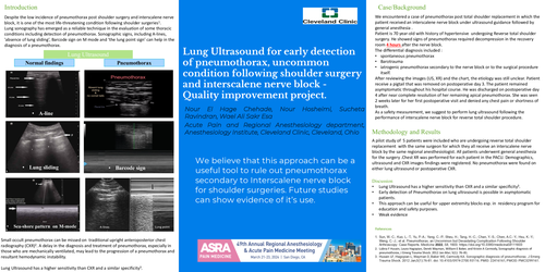
Despite the low incidence of pneumothorax post-shoulder surgery and interscalene nerve block, it is one of the most life-threatening condition following shoulder surgeries1.Lung sonography has emerged as a reliable technique in the evaluation of some thoracic conditions including detection of pneumothorax. Sonographic signs, including A-lines, 'absence of lung sliding’, Barcode sign on M mode and ‘the lung point sign’ can help in the diagnosis of a pneumothorax. Small occult pneumothorax can be missed on traditional upright anteroposterior chest radiography (CXR)2. A delay in the diagnosis and treatment of pneumothorax, especially in those who are mechanically ventilated, may lead to the progression of a pneumothorax and resultant hemodynamic instability.Lung Ultrasound has a higher sensitivity than CXR and a similar specificity3.
Case/Background
We encountered a case of pneumothorax post total shoulder replacement in which the patient received an interscalene nerve block under ultrasound guidance followed by general anesthesia .
Patient is 70 year-old with history of hypertensive undergoing Reverse total shoulder surgery. He showed signs of pneumothorax required decompression in the recovery room 4 hours after the nerve block.
The differential diagnosis included :
After reviewing the images (US, XR) and the chart, the etiology was still unclear. Patient receive a pigtail that was removed on postoperative day 3. The patient remained asymptomatic throughout his hospital course. He was discharged on postoperative day 4 after near complete resolution of her remaining apical pneumothorax. She was seen 2 weeks later for her first postoperative visit and denied any chest pain or shortness of breath.
As a safety measurement, we suggest to perform lung ultrasound following the performance of interscalene nerve block for reverse total shoulder procedure
Methodology and results
A pilot study of 5 patients were included who are undergoing reverse total shoulder replacement with the same surgeon for which they all receive an interscalene nerve block by the same regional anesthesiologist. All patients underwent general anesthesia for the surgery .Chest XR was performed for each patient in the PACU. Demographics, ultrasound and CXR images findings were registered. No pneumothorax were found on either lung ultrasound or postoperative CXR