457 posters, 2 videos, 2 audios, 8 topics, 45 sessions, 401 authors, 265 institutions
ePostersLive by SciGen Technologies S.A. All rights reserved.
49th Annual Regional Anesthesiology and Acute Pain Medicine Meeting
March 21-23, 2024 | The Hilton Bayfront San Diego | San Diego, California

5437
DETERMINATION OF SAFE DISCONTINUATION OF EPIDURAL CATHETER IN LUNG TRANSPLANT PATIENT FOLLOWING EMERGENT INITIATION OF VV-ECMO
Poster Presenter
Authors
Affiliations
Part of Topic
Medically Challenging Cases
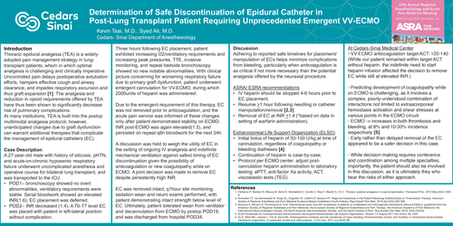
Determination of Safe Discontinuation of Epidural Catheter in Post-Lung Transplant Patient Requiring Unprecedented Emergent VV-ECMO
Kevin Tsai, M.D., Syed Ali, M.D.
Cedars- Sinai Department of Anesthesiology
Introduction
Thoracic epidural analgesia (TEA) is a widely-adopted pain management strategy in lung-transplant patients, whom in which optimal analgesia is challenging and clinically imperative. Uncontrolled pain delays postoperative extubationefforts, hampers effective cough and airway clearance, and impedes respiratory excursion and thus graft expansion [1]. The analgesia and reduction in opioid requirements offered by TEA have thus been shown to significantly decrease risk of pulmonary complications.
At many institutions, TEA is built into the postop multimodal analgesia protocol; however, unanticipated changes due to graft dysfunction can warrant additional therapies that complicate the management of epidural catheters (EC).
Case Description
A 27-year-old male with history of silicosis, pHTN, and acute-on-chronic hypoxemic respiratory failure underwent uncomplicated anesthesia and operative course for bilateral lung transplant, and was transported to the ICU.
Three hours following EC placement, patient exhibited increasing O2/ventilatory requirements and increasing peak pressures. TTE, invasive monitoring, and repeat bedside bronchoscopy showed no new notable abnormalities. With clinical picture concerning for worsening respiratory failure due to primary graft dysfunction, patient underwent emergent cannulation for VV-ECMO, during which 2000units of heparin was administered.
Due to the emergent requirement of this therapy, EC was not removed prior to anticoagulation, and the acute pain service was informed of these changes only after patient demonstrated stability on ECMO. INR post-ECMO was again elevated(1.5), and persisted on repeat q6h bloodwork for the next 24h.
A discussion was held to weigh the utility of EC in the setting of ongoing IV analgesia and indefinite mechanical ventilation against safest timing of EC discontinuation given the possibility of anticoagulation or new coagulopathy while on ECMO. A joint decision was made to remove EC despite persistently high INR.
EC was removed intact; q1hour site monitoring, sedation wean and neuro exams performed, with patient demonstrating intact strength below level of EC. Ultimately, patient tolerated wean from ventilator and decannulation from ECMO by postop POD19, and was discharged from hospital POD34.
Discussion
Adhering to reported safe timelines for placement/ manipulation of ECs helps minimize complications from bleeding, particularly when anticoagulation is as critical if not more necessary than the potential analgesia offered by the neuraxial procedure.
ASRA/ ESRA recommendations
Extracorporeal Life Support Organization (ELSO)
At Cedars-Sinai Medical Center
• VV-ECMO anticoagulation target ACT: 120-140
(While our patient remained within target ACT without heparin, the indefinite need to start heparin infusion affected the decision to remove EC while still at elevated INR.)
- Predicting development of coagulopathy while on ECMO is challenging, as it involves a complex, poorly-understood combination of interactions not limited to extraacorporealhemostasis activation and shear stresses at various points in the ECMO circuit.
- ECMO -> increases in both thrombosis and bleeding, at 8% and 10-30% incidence respectively [5].
- Early rather than delayed removal of the EC appeared to be a safer decision in this case.
- While decision-makingrequires conference and coordination among multiple specialties, importantly, the patient should also be involved in this discussion, as it is ultimately they who bear the risks of either approach.