457 posters, 2 videos, 2 audios, 8 topics, 45 sessions, 401 authors, 265 institutions
ePostersLive by SciGen Technologies S.A. All rights reserved.
49th Annual Regional Anesthesiology and Acute Pain Medicine Meeting
March 21-23, 2024 | The Hilton Bayfront San Diego | San Diego, California

5131
EVALUATION OF HYPOTENSION AFTER THORACIC EPIDURAL PLACEMENT
Poster Presenter
Authors
Affiliations
Part of Topic
Medically Challenging Cases
Background
Rib fractures are often present in the trauma population and the resulting thoracic pain can cause impaired respiratory effort, which increases the risk of respiratory compromise. Thoracic epidural analgesia (TEA) provides superior pain relief compared to other modalities.1 However, due to the blind nature of TEA placement, malposition can occur despite adequate localization, smooth catheter threading and negative aspiration and test dose. Therefore, hypotension after TEA placement warrants thorough investigation to evaluate for possible intrathecal, intravascular, or intrapleural placement.2 This case report describes an incidentally found intrapleural thoracic epidural in a patient with multiple rib and extremity fractures.
Case Description
Patient is a 76-year-old female (147cm, BMI 23) with hypertension, insomnia, osteoporosis, migraines, and papillary thyroid cancer who presented to the hospital after a motor vehicle accident. Her injuries included fractures in right ribs 1-8, left ribs 2-6, and left ribs 10-12 as well as multiple extremity fractures. Initial CT scans did not show pneumothorax and patient was saturating above 94% on 2L nasal cannula. The acute pain service was consulted for TEA.
Patient was placed in the left lateral decubitus position. A paramedian approach was used with a 17-gauge, 3.5 inch-Tuohy needle to enter the posterior epidural space at T4-5. Loss of resistance to saline was met at 6cm. Then, a catheter was easily threaded. The needle and stylet were removed intact and the catheter was pulled back to 12 cm hashmarks at the skin leaving 6cm of catheter in the presumed epidural space. After negative aspiration, 3ml test dose of 2% lidocaine with epinephrine (1:200,000) was administered and was again negative. The catheter was secured, and a patient controlled epidural anesthesia was initiated with hydromorphone 12.5mg/ml and bupivacaine 0.0625% at 4ml/hour, 2ml bolus every 10 minutes, and 4ml clinician bolus.
Patient’s pain numerical rating scale improved from 8 to 6. However, 30 minutes after placement, patient experienced hypotension with mean arterial pressure of 48 mm Hg from 81 and was started on a norepinephrine infusion up to 5mcg/min. Concurrently, patient labs also showed a hemoglobin drop from 10.3g/dl to 9.3 and was transfused one unit packed red blood cells. After the transfusion, patient was able to be weaned off of norepinephrine.
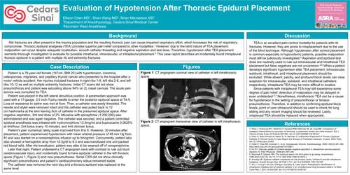
Later that night, Patient underwent a CT angiogram neck with contrast to rule out blunt cerebrovascular injury, and incidentally found to have epidural catheter in the left thoracic space (Figure 1, Figure 2) and new pneumothorax. Serial CXR did not show clinically significant pneumothorax and patient’s cardiopulmonary status remained stable.
The catheter was removed the next day and a thoracic epidural was replaced at the same level.
Discussion
TEA is an excellent pain control modality for patients with rib fractures. However, they are prone to misplacement due to the use of the blind technique. Although hypotension after correct placement is common especially in hypovolemic patients, possible malposition must still be judiciously investigated.3 Aspiration and epidural test dose are routinely used to rule out intravascular and intrathecal TEA placement but false negatives are not uncommon.4,5 When a patient develops significant hypotension after TEA placement, intravascular, subdural, intrathecal, and intrapleural placement should be excluded. While absent, patchy, and profound block levels can raise suspicion for intravascular, subdural, and intrathecal catheters respectively, intrapleural TEA may be more difficult to localize.
Since patients with intrapleural TEA may still experience some degree of pain relief, detection of malposition may be delayed or even undetected.6,7 Nonetheless, intrathoracic TEA can be a cause for hypotension in the setting of pneumothorax or tension pneumothorax. Therefore, in addition to confirming epidural block levels, point of care ultrasound should be used to check for lung sliding and any recent imaging should be reviewed. Lastly, misplaced TEA should be replaced when appropriate.