457 posters, 2 videos, 2 audios, 8 topics, 45 sessions, 401 authors, 265 institutions
ePostersLive by SciGen Technologies S.A. All rights reserved.
49th Annual Regional Anesthesiology and Acute Pain Medicine Meeting
March 21-23, 2024 | The Hilton Bayfront San Diego | San Diego, California

5458
ULTRASOUND-ASSISTED SPINAL ANESTHESIA IN SETTING OF POST-COVID PULMONARY SEQUELAE
Poster Presenter
Authors
Affiliations
Part of Topic
Medically Challenging Cases
Ultrasound-Assisted Spinal Anesthesia in the Setting of Post-COVID Pulmonary Sequelae
Jacy Gressen, M.D., Alexander Reskallah, M.D., Hart Donahue, M.D., Evan Wright, M.D., Stephanie L. Bradley M.D., M.P.H, Emily A. Lai, M.D., Andrzej P. Kwater, M.D. Department of Anesthesiology and Perioperative Medicine, The University of Texas MD Anderson Cancer Center, Houston, TX
Introduction: Injection of local anesthesia into the intrathecal space can provide effective surgical anesthesia for abdominal and lower extremity surgeries. In patients with pulmonary comorbidities, elevated risks for post-intubation morbidity increases the appeal for spinal anesthesia as the primary anesthetic plan. Patient body habitus can complicate placement of spinal anesthesia by traditional manual identification of palpable surface anatomy. Here, we describe ultrasound guidance for successful placement of spinal anesthetic in a severely obese patient with significant post-COVID pulmonary sequelae.
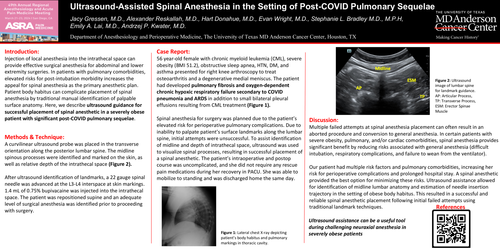
Methods & Technique: A curvilinear ultrasound probe was placed in the transverse orientation along the posterior lumbar spine. The midline spinous processes were identified and marked on the skin, as well as relative depth of the intrathecal space (Figure 2). After ultrasound identification of landmarks, a 22 gauge spinal needle was advanced at the L3-L4 interspace at skin markings. 1.4 mL of 0.75% bupivacaine was injected into the intrathecal space. The patient was repositioned supine and an adequate level of surgical anesthesia was identified prior to proceeding with surgery.
Case Report: 56 year-old female with chronic myeloid leukemia (CML), severe obesity (BMI 51.2), obstructive sleep apnea, HTN, DM, and asthma presented for right knee arthroscopy to treat osteoarthritis and a degenerative medial meniscus. The patient had developed pulmonary fibrosis and oxygen-dependent chronic hypoxic respiratory failure secondary to COVID pneumonia and ARDS in addition to small bilateral pleural effusions resulting from CML treatment (Figure 1). Spinal anesthesia for surgery was planned due to the patient’s elevated risk for perioperative pulmonary complications. Due to inability to palpate patient’s surface landmarks along the lumbar spine, initial attempts were unsuccessful. To assist identification of midline and depth of intrathecal space, ultrasound was used to visualize spinal processes, resulting in successful placement of a spinal anesthetic. The patient’s intraoperative and postop course was uncomplicated, and she did not require any rescue pain medications during her recovery in PACU. She was able to mobilize to standing and was discharged home the same day.
Discussion: Multiple failed attempts at spinal anesthesia placement can often result in an aborted procedure and conversion to general anesthesia. In certain patients with severe obesity, pulmonary, and/or cardiac comorbidities, spinal anesthesia provides significant benefit by reducing risks associated with general anesthesia (difficult intubation, respiratory complications, and failure to wean from the ventilator). Our patient had multiple risk factors and pulmonary comorbidities, increasing her risk for perioperative complications and prolonged hospital stay. A spinal anesthetic provided the best option for minimizing these risks. Ultrasound assistance allowed for identification of midline lumbar anatomy and estimation of needle insertion trajectory in the setting of obese body habitus. This resulted in a successful and reliable spinal anesthetic placement following initial failed attempts using traditional landmark techniques.
Ultrasound assistance can be a useful tool during challenging neuraxial anesthesia in severely obese patients.