457 posters, 2 videos, 2 audios, 8 topics, 45 sessions, 401 authors, 265 institutions
ePostersLive by SciGen Technologies S.A. All rights reserved.
49th Annual Regional Anesthesiology and Acute Pain Medicine Meeting
March 21-23, 2024 | The Hilton Bayfront San Diego | San Diego, California

5195
A NOVEL APPROACH TO THE CORDS OF THE BRACHIAL PLEXUS TO DECREASE THE RISK OF PULMONARY COMPROMISE
Poster Presenter
Authors
Affiliations
Part of Topic
Medically Challenging Cases
Title: A Fascial Plane Approach to the Cords of the Brachial Plexus
Authors: Emma Rousakis, MD, Jacob Whittle, MD, Alastair E. Moody, MD, Evelyn Loose, MD, Jeffrey Swenson, MD
Abstract:
Introduction: Brachial plexus block for analgesia of the elbow and distal humerus is commonly performed at the supraclavicular or infraclavicular level (1). Ultrasound guided nerve blocks at these sites are effective but require injection in proximity to major vascular structures and nerves of the brachial plexus (2,3). Moreover, both techniques can be associated with pneumothorax and diaphragm dysfunction (4,5).
We present a fascial plane technique for nerve block at the level of the cords of the brachial plexus. This block is performed by injection into the tissue plane separating the coracobrachialis and subscapularis muscles. Injection at this level is proximal enough to provide analgesia of the elbow and distal humerus while avoiding the risk of pneumothorax or diaphragm dysfunction. The injection can be performed within a tissue plane 1 cm superior to the neurovascular structures.
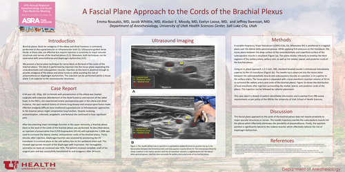
Materials & Methods: A variable frequency, linear transducer (LOGIQ E10s, GE, Milwaukee WI) is positioned in a sagittal plane over the lateral delto-pectoral groove. While applying firm pressure on the transducer, the tissue plane between the deep surface of the coracobrachialis (CM) and superficial surface of the subscapularis muscle (SM) is visualized (Figure 1a). This plane divides inferiorly to envelop the third segment of the axillary artery, axillary vein, as well as the medial, lateral, and posterior cords of the brachial plexus.
Using an in-plane approach, a 2.5 inch, 18G, standard beveled needle is introduced immediately superior to the US transducer (Figure 1b). The needle tip is advanced into the tissue plane between the coracobrachialis muscle and subscapularis muscles at a position 1 cm superior to the axillary artery. The tissue plane is expanded with a local anesthetic injection volume of 30 ml to surround the axillary artery and cords of the brachial plexus. Figure 1b shows the distribution of local anesthetic after injection surrounding the medial, lateral, and posterior cords of the plexus. This injection can be followed by catheter placement.
This case report is devoid of patient identifiable information and is exempt from IRB review requirements as per policy of the IRB for the University of Utah School of Health Sciences.
Case Report:
A 64 year old, 45kg, 163 cm female with pseudotumor of the elbow was treated surgically with extensive debridement of the distal humerus and excision of the radial head. In the PACU, she experienced severe postoperative pain in the elbow and distal humerus. Her past medical history of chronic lung disease and cervical spine fusion made effective analgesia difficult since traditional supraclavicular or infraclavicular approaches to the brachial plexus might compromise lung function. Despite receiving acetaminophen, celecoxib, pregabalin, and fentanyl she continued to have significant pain.
After documenting intact neurologic function in the upper extremity, a brachial plexus block at the level of the cords of the brachial plexus was performed. As described above, an injection of preservative free 0.25% bupivacaine (30 ml) with epinephrine 1:200k was used to surround the lateral, medial, and posterior cords of the brachial plexus. Thirty minutes after injection, diaphragm function was assessed by positioning the US transducer in a coronal plane at the mid-axillary line on the ipsilateral chest wall. This showed appropriate descent of the diaphragm with inspiration. Her hemoglobin saturation on room air remained over 95%. The patient received complete relief of her surgical pain and was successfully transitioned to oral analgesics after 24 hours.
Discussion: This fascial plane approach to the cords of the brachial plexus does not require proximity to major vascular structures or nerves. The needle trajectory overlies the subscapularis muscle not the pleura which effectively eliminates the possibility of pneumothorax. Finally, the injectate position is significantly lateral to the scalene muscles which effectively reduces the risk of diaphragm dysfunction.
References
1.Feigl GC, Litz RJ, Marhofer P. Anatomy of the brachial plexus and its implications for daily clinical practice: regional anesthesia is applied anatomy..Reg Anesth Pain Med. 2020 Aug;45(8):620-627.
2. Boivin A, Nadeau MJ, Dion N, Lévesque S, Nicole PC, Turgeon AF. Ultrasound-Guided Single-Injection Infraclavicular Block Versus Ultrasound-Guided Double-Injection Axillary Block: A Noninferiority Randomized Controlled Trial. Anesth Analg. 2016 Jan;122(1):273-8
3.Hanumanthaiah D, Vaidiyanathan S, Garstka M, Szucs S. Iohom G. Ultrasound guided supraclavicular block..Med Ultrason. 2013 Sep;15(3):224-9
4. Abell DJ, Barrington MJ. Pneumothorax after ultrasound-guided supraclavicular block: presenting features,risk,and related training. Reg Anesth Pain Med. 2014 Mar-Apr;39(2):164-7
5. Park SK, Lee SY, Kim WH, Park HS, Lim YJ, Bahk JH.Comparison of Supraclavicular and Infraclavicular Brachial Plexus Block: A Systemic Review of Randomized Clinical Trials. Anesth Analg. 2017 Feb;124(2):636-644