457 posters, 2 videos, 2 audios, 8 topics, 45 sessions, 401 authors, 265 institutions
ePostersLive by SciGen Technologies S.A. All rights reserved.
49th Annual Regional Anesthesiology and Acute Pain Medicine Meeting
March 21-23, 2024 | The Hilton Bayfront San Diego | San Diego, California

5193
DIAGNOSTIC ULTRASOUND-GUIDED REGIONAL BLOCKADE TO FACILITATE PERIPHERAL SCHWANNOMA RESECTION
Poster Presenter
Authors
Affiliations
Part of Topic
Medically Challenging Cases
Introduction:
The application for ultrasound-guided regional nerve blocks is constantly expanding.
At our institution, diagnostic regional nerve blocks have faciliated surgical planning, particularly for neuropathologies such as nerve tumors and chronic neuropathic pain.
Schwannomas are the most common neoplasm affecting peripheral nerves.1 Surgical resection is the standard of care; however, resection carries the risk of potential nerve injury.2 We present a case where ultrasound-guided diagnostic blockade of peripheral schwannomas was utilized to localize the source of a patient’s neuropathic pain and to plan for surgical management with potential for targeted resection.
Material & Methods:
Informed consent for inclusion in this case report was obtained. As this case report is devoid of any patient identifiable information, it is exempt from IRB review requirements per the MD Anderson Cancer Center policy.
Case Report:
65 year-old woman with a history of recurrent breast cancer, MEN2a syndrome, and multiple peripheral schwannomas s/p prior excision presented with persistent neuropathic pain due to several lower extremity schwannomas.
After being seen in clinic, neurosurgery consulted the acute pain service to perform diagnostic blocks of the identified schwannomas to determine if blockade with local anesthetic resulted in analgesia and helped to locate the most prominent lesion to target for resection.
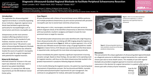
The well-demarcated schwannomas were easily identified with a high-frequency linear ultrasound probe after correlating with MRI imaging along the trajectory of the superficial peroneal nerve (Figure 1). With an in-plane approach, 3-4 cc of 2% lidocaine was infiltrated around each lesion using a 23 gauge hypodermic needle (Figure 2). A total of 10 mL of 2% lidocaine was injected around the three lesions in sequential order, starting with the most distal and progressing proximally.
Almost immediately the patient reported a significant improvement in burning sensation and gait. Based on these findings, she was taken to the operating room for targeted resection, with focus on the distal schwannomas that resulted in the greatest improvement in symptoms following diagnostic blockade.
In the 6 months following the targeted resection, the patient has reported gradual improvement of dysesthesias and pain. She has discontinued NSAIDs, tramadol, and hydrocodone/acetaminophen requirements for pain management. She continues to use gabapentin for residual, attenuated neuropathic pain.
Discussion:
This case presents the unique application of ultrasound-guided regional blockade as a diagnostic precursor to definitive surgical intervention for a patient with chronic pain due to nerve sheath tumors. This modality of care with regional blockade also provided a targeted approach to resection and provided reassurance in the decision for surgical intervention in a patient suffering from chronic pain with a history of prior resections.