457 posters, 2 videos, 2 audios, 8 topics, 45 sessions, 401 authors, 265 institutions
ePostersLive by SciGen Technologies S.A. All rights reserved.
49th Annual Regional Anesthesiology and Acute Pain Medicine Meeting
March 21-23, 2024 | The Hilton Bayfront San Diego | San Diego, California

5262
ULTRASOUND-GUIDED SCALP BLOCK FOR AN AWAKE CRANIOTOMY
Poster Presenter
Authors
Affiliations
Part of Topic
Medically Challenging Cases
Ultrasound-guided Scalp Block
for an
Awake Craniotomy
Carlos G. Montero, MD, Fernando Vazquez, MD, Richard Marks, MD and
Jonathan Metry, MD
INTRODUCTION
Awake craniotomies allow neurosurgeons to achieve
a higher rate of brain tumor resection while minimizing
significant neurological deficits. Anesthesiologists can
play a crucial role by performing scalp blocks, which
include seven bilateral nerves, implementing a sedative
plan that maintains a patent airway and a plan that
reduces interference with intraoperative neuromonitoring.
Awake craniotomies reduce the side effects of general
anesthesia, promote early postoperative neurological
evaluation, and improve resource utilization.1
In this case report, we present a patient who
underwent an awake craniotomy for a frontotemporaloccipital
brain tumor.
PATIENT PRESENTATION
Our patient is a 74-year-old female (weight 100 kg
and BMI 34) with significant past medical history of
hypertension on metoprolol, aortic prosthetic mechanical
valve on Warfarin, trigeminal neuralgia, and left temporooccipital
brain tumor associated with gait imbalance and
memory and word-finding deficits. She presented for
awake left-sided craniotomy with intraoperative brain
mapping. Airway examination demonstrated Mallampati 2,
thyromental distance >3 fingerbreadths, and full range of
motion of the neck. An asleep-awake-asleep technique
was implemented using general anesthesia with invasive
monitoring, monitored anesthesia care, and regional
anesthesia.
Standard ASA monitors were placed, and a time out
was performed. After an uneventful induction and
intubation, dexmedetomidine, propofol, and remifentanil
infusions were started.
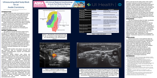
Ultrasound guided bilateral scalp blocks and
superficial cervical plexus blocks were performed. The
patient was positioned in the supine position and a total of
25 cc of 0.5% Bupivacaine was injected under ultrasound
guidance. Bilateral supraorbital, supratrochlear,
zygomaticotemporal, and greater occipital, nerves were
individually blocked and the greater auricular and lesser
occipital nerves were blocked via the superficial cervical
plexus. A single intravenous dose of dexamethasone was
given as an adjuvant to prolong the duration and improve
the quality of analgesia provided by the ultrasound-guided
scalp blocks.2
After completion of the ultrasound-guided nerve blocks,
the patient was positioned for surgery and placed in pins
with no change in vital signs. Additionally, vital signs
remained unchanged following surgical incision with no
adjustments to the propofol, remifentanil, and
dexmedetomidine infusions.
When it was time for the awake portion of the surgery,
the patient underwent successful emergence and
extubation. Awake language mapping was performed to
maximize extent of resection and minimize functional
deficits. The patient was cooperative, calm and pain-free
throughout the awake portion of the procedure. At the
conclusion of the awake portion of the surgery, the
patient was induced and reintubated. Final motor
analysis was performed, and the patient was extubated
and transferred to the neurosurgical intensive care unit
without complications. In the immediate post-operative
period, the patient’s pain scores ranged from 0-2/10 and
required no additional analgesics.
DISCUSSION
The use of ultrasound allows for better visualization
of anatomy and neurovascular distribution, in
comparison to landmark-based scalp blocks, and may
reduce volume and concentration of local anesthetic
needed for an adequate block, thus reducing the risk of
local anesthetic systemic toxicity (LAST).3 Complications
from scalp blocks may include infection, hemodynamic
changes and arrythmias if epinephrine is used, vascular
injury and hematoma, unilateral ptosis, facial nerve
palsy, bradycardia and hypotension from the
trigeminocardiac reflex, intracerebral or subarachnoid
injection, anaphylactic response to local anesthetic, and
LAST.4
CONCLUSION
Postcraniotomy pain is most significant within the
first 48 hours post op and presents as a headache that
is generalized and pounding or pulsatile. The pain is
mostly superficial suggesting somatic rather than
visceral pain.6 This case report demonstrates that
ultrasound-guided scalp blocks provide the analgesia
required to perform an awake craniotomy. A
multidisciplinary team can interact with the awake
patient with minimal discomfort and hemodynamic
instability during pin application and scalp incision.
Regional anesthesia can play an important role to
reduce opioid requirements and allow for optimal
conditions for a neurological assessment.
REFERENCES
1. Amiri HR, Kouhnavard M, Safari S. Scalp block for awake craniotomy in a patient
with a frontal bone mass: a case report. Anesth Pain Med. 2012 Winter;1(3):187-90.
doi: 10.5812/kowsar.22287523.3608. Epub 2012 Jan 1
2. Zhao C, Jia Z, Shrestha N, Luo F. REDUCE trial: the effects of perineural
dexamethasone on scalp nerve blocks for relief of postcraniotomy pain-a study
protocol for a randomized controlled trial. Trials. 2021 Nov 4;22(1):772. doi:
10.1186/s13063-021-05747-y. PMID: 34736497; PMCID: PMC8567555.
3. Archer DP, McKenna JM, Morin L, Ravussin P. Conscious-sedation analgesia during
craniotomy for intractable epilepsy: a review of 354 consecutive cases. Can J
Anaesth. 1988;35:338–344.
4. Supriya Dsouza, Darreul Sewell. Scalp Block: Technique and Applications. World
Federation of Societies of Anesthesiologists.
5. Cally Burnand, Joseph Sebastian, Anaesthesia for awake craniotomy, Continuing
Education in Anaesthesia Critical Care & Pain, Volume 14, Issue 1, February 2014,
Pages 6–11
6. De Benedittis G, Lorenzetti A, Migliore M, et al. Postoperative pain in
neurosurgery: a pilot study in brain surgery. Neurosurgery. 1996;38(3):466–9;
discussion 469–70.
7. Paul J. Zetlaoui, Elodie Gauthier, Dan Benhamou, Ultrasound-guided scalp nerve
blocks for neurosurgery: A narrative review, Anaesthesia Critical Care & Pain
Medicine, Volume 39, Issue 6, 2020, Pages 876-882.
8. International Anesthesiology Clinics62(1):10-20, Winter 2024.