501 posters, 2 videos, 1 audios, 8 topics, 37 sessions, 395 authors, 307 institutions
ePostersLive by SciGen Technologies S.A. All rights reserved.
23rd Annual Pain Medicine Meeting 2024
November 21-23, 2024 | Caesars Palace, Las Vegas, Nevada

6012
AN UNUSUAL PAIN MANAGEMENT DIAGNOSIS: INCIDENTAL DISCOVERY OF LOW-GRADE GLIOMA IN A PATIENT WITH CERVICOTHORACIC PAIN
Poster Presenter
Part of Topic
Medically Challenging Cases
An Unusual Pain Management Diagnosis: Incidental discovery of low-grade glioma in a patient with cervicothoracic pain
Ryan Shields MD (1) and Gwynne Kirchen MD (2)
(1) Department of Physical Medicine and Rehabilitation, (2) Department of Anesthesiology - Pain Medicine Division, Medical College of Wisconsin
Case Description
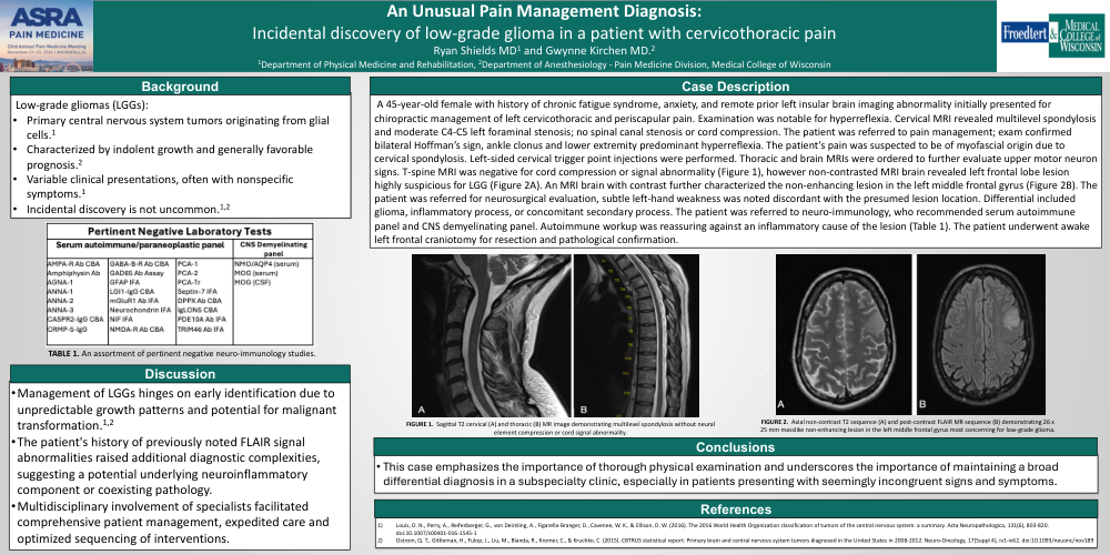
A 45-year-old female with history of chronic fatigue syndrome, anxiety, and remote prior left insular brain imaging abnormality initially presented for chiropractic management of left cervicothoracic and periscapular pain. Examination was notable for hyperreflexia. Cervical MRI revealed multilevel spondylosis and moderate C4-C5 left foraminal stenosis; no spinal canal stenosis or cord compression. The patient was referred to pain management; exam confirmed bilateral Hoffman’s sign, ankle clonus and lower extremity predominant hyperreflexia. The patient’s pain was suspected to be of myofascial origin due to cervical spondylosis. Left-sided cervical trigger point injections were performed. Thoracic and brain MRIs were ordered to further evaluate upper motor neuron signs. T-spine MRI was negative for cord compression or signal abnormality (Figure 1), however non-contrasted MRI brain revealed left frontal lobe lesion highly suspicious for LGG (Figure 2A). An MRI brain with contrast further characterized the non-enhancing lesion in the left middle frontal gyrus (Figure 2B). The patient was referred for neurosurgical evaluation, subtle left-hand weakness was noted discordant with the presumed lesion location. Differential included glioma, inflammatory process, or concomitant secondary process. The patient was referred to neuro-immunology, who recommended serum autoimmune panel and CNS demyelinating panel. Autoimmune workup was reassuring against an inflammatory cause of the lesion (Table 1). The patient underwent awake left frontal craniotomy for resection and pathological confirmation.