501 posters, 2 videos, 1 audios, 8 topics, 37 sessions, 395 authors, 307 institutions
ePostersLive by SciGen Technologies S.A. All rights reserved.
23rd Annual Pain Medicine Meeting 2024
November 21-23, 2024 | Caesars Palace, Las Vegas, Nevada

5984
AN UNUSUAL CASE OF AN OSTEOID OSTEOMA IN THE THORACIC SPINE: A CASE REPORT AND LITERATURE REVIEW
Poster Presenter
Part of Topic
Medically Challenging Cases
Osteoid osteoma (OO) is a benign bone tumor that does not have malignant potential.1 It is generally small and usually affects young males 5-30 years old.2 OO accounts for 10-14% of all benign bone tumors and most often occurs in the long bones. Approximately 6-20% of osteoid osteomas occur in the spine, with only 12% of these occurring in the thoracic region.2 While a portion of the spinal osteoid osteomas may present with painful secondary scoliosis, those that are located in the cervical spine more often present with a painful, restricted range of motion.3,4 The natural course of this disease varies greatly, and the severity of symptoms often depends on location. While many treatment modalities are available, there are reports of osteoid osteomas regressing spontaneously in six years' time with conservative treatment.4
-
24-year-old male with gradual, worsening pain in spine and right upper shoulder blade for one year after an extended period of weight training
-
Initial plain radiography and MRI were unremarkable and the working diagnosis was myofascial pain syndrome vs chronic muscle overuse
-
Initial treatment regimen consisted of conservative management with NSAIDs, acetaminophen, physical therapy, and acupuncture
-
After failing conservative management, trigger points, and an erector spinae block were performed without sustained relief.
-
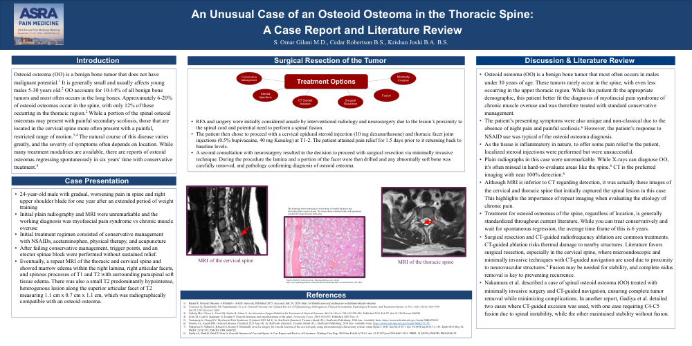
Eventually, a repeat MRI of the thoracic and cervical spine and showed marrow edema within the right lamina, right articular facets, and spinous processes of T1 and T2 with surrounding paraspinal soft tissue edema. There was also a small T2 predominantly hypointense, heterogenous lesion along the superior articular facet of T2 measuring 1.1 cm x 0.7 cm x 1.1 cm, which was radiographically compatible with an osteoid osteoma.
-
RFA and surgery were initially considered unsafe by interventional radiology and neurosurgery due to the lesion’s proximity to the spinal cord and potential need to perform a spinal fusion.
-
The patient then chose to proceed with a cervical epidural steroid injection (10 mg dexamethasone) and thoracic facet joint injections (0.5% bupivacaine, 40 mg Kenalog) at T1-2. The patient attained pain relief for 1.5 days prior to it returning back to baseline levels.
-
A second consultation with neurosurgery resulted in the decision to proceed with surgical resection via minimally invasive technique. During the procedure the lamina and a portion of the facet were then drilled and any abnormally soft bone was carefully removed, and pathology confirming diagnosis of osteoid osteoma.
-
Osteoid osteoma (OO) is a benign bone tumor that most often occurs in males under 30 years of age. These tumors rarely occur in the spine, with even less occurring in the upper thoracic region. While this patient fit the appropriate demographic, this patient better fit the diagnosis of myofascial pain syndrome of chronic muscle overuse and was therefore treated with standard conservative management.
-
The patient’s presenting symptoms were also unique and non-classical due to the absence of night pain and painful scoliosis.6 However, the patient’s response to NSAID use was typical of the osteoid osteoma diagnosis.
-
As the tissue is inflammatory in nature, to offer some pain relief to the patient, localized steroid injections were performed but were unsuccessful.
-
Plain radiographs in this case were unremarkable. While X-rays can diagnose OO, it's often missed in hard-to-evaluate areas like the spine.6 CT is the preferred imaging with near 100% detection.6
-
Although MRI is inferior to CT regarding detection, it was actually these images of the cervical and thoracic spine that initially captured the spinal lesion in this case. This highlights the importance of repeat imaging when evaluating the etiology of chronic pain.
-
Treatment for osteoid osteomas of the spine, regardless of location, is generally standardized throughout current literature. While you can treat conservatively and wait for spontaneous regression, the average time frame of this is 6 years.
-
Surgical resection and CT-guided radiofrequency ablation are common treatments. CT-guided ablation risks thermal damage to nearby structures. Literature favors surgical resection, especially in the cervical spine, where microendoscopic and minimally invasive techniques with CT-guided navigation are used due to proximity to neurovascular structures.6 Fusion may be needed for stability, and complete nidus removal is key to preventing recurrence.
-
Nakamura et al. described a case of spinal osteoid osteoma (OO) treated with minimally invasive surgery and CT-guided navigation, ensuring complete tumor removal while minimizing complications. In another report, Gadiya et al. detailed two cases where CT-guided excision was used, with one case requiring C4-C5 fusion due to spinal instability, while the other maintained stability without fusion.