501 posters, 2 videos, 1 audios, 8 topics, 37 sessions, 395 authors, 307 institutions
ePostersLive by SciGen Technologies S.A. All rights reserved.
23rd Annual Pain Medicine Meeting 2024
November 21-23, 2024 | Caesars Palace, Las Vegas, Nevada

6237
ACETABULAR CARCINOMA IN A PATIENT EXPERIENCING SUSPECTED RADICULAR LOW BACK PAIN
Poster Presenter
Authors
Affiliations
Part of Topic
Medically Challenging Cases
Introduction
•Worldwide, low back pain (LBP) is the single leading cause of disability and need for physical rehabilitation, with nonspecific and insidious back pain comprising most clinical cases.
•As the general population continues to live longer, pain physicians will see an increased prevalence of both typical and atypical presentations of low back pain in their clinics.
•This case was written with patient consent.
Case
•Patient: KP, 60-year-old Caucasian female, pertinent history of R tibial fracture repair secondary to mechanical fall in 2014. No known oncologic history.
•
•Starting in August 2019, Patient began to experience lower back pain with involvement of her right lower limb. After an L3/L4 transforaminal epidural steroid injection at an outside hospital provided no relief, she presented to the University of Illinois ED, where she was referred to our pain clinic for management.
•Patient presented to our clinic with similar symptoms; she endorsed a four-month history of worsening radiating pain down her right leg with increased severity in her upper thigh and radiation into her toes with numbness and tingling but denied incontinence, saddle anesthesia, or systemic symptoms.
•We ordered an MRI of the hip, which showed a malignant pathology of the hip region not seen on prior spinal imaging.
•Given our findings of hip pathology versus spinal etiology, she was referred to UIC orthopedics clinic for further workup, where she ultimately endorsed a month-long history of progressive inability to bear weight, nocturnal pain, and unintentional 20 lb. weight loss.
Diagnostic Imaging
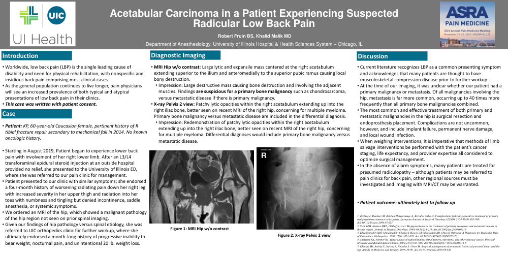
•MRI Hip w/o contrast: Large lytic and expansile mass centered at the right acetabulum extending superior to the ilium and anteromedially to the superior pubic ramus causing local bony destruction.
•Impression: Large destructive mass causing bone destruction and involving the adjacent muscles. Findings are suspicious for a primary bone malignancy such as chondrosarcoma, versus metastatic disease if there is primary malignancy.
•X-ray Pelvis 2 view: Patchy lytic opacities within the right acetabulum extending up into the right iliac bone, better seen on recent MRI of the right hip, concerning for multiple myeloma. Primary bone malignancy versus metastatic disease are included in the differential diagnosis.
•Impression: Redemonstration of patchy lytic opacities within the right acetabulum extending up into the right iliac bone, better seen on recent MRI of the right hip, concerning for multiple myeloma. Differential diagnoses would include primary bone malignancy versus metastatic disease.
Discussion
•Current literature recognizes LBP as a common presenting symptom and acknowledges that many patients are thought to have musculoskeletal compression disease prior to further workup.
•At the time of our imaging, it was unclear whether our patient had a primary malignancy or metastasis. Of all malignancies involving the hip, metastasis is far more common, occurring up to 40 times more frequently than all primary bone malignancies combined.
•The most common and effective treatment of both primary and metastatic malignancies in the hip is surgical resection and endoprosthesis placement. Complications are not uncommon, however, and include implant failure, permanent nerve damage, and local wound infection.
•When weighing interventions, it is imperative that methods of limb salvage interventions be performed with the patient’s cancer staging, life expectancy, and provider expertise all considered to optimize surgical management.
•In the absence of alarm symptoms, many patients are treated for presumed radiculopathy – although patients may be referred to pain clinics for back pain, other regional sources must be investigated and imaging with MRI/CT may be warranted.
•Patient outcome: ultimately lost to follow up