501 posters, 2 videos, 1 audios, 8 topics, 37 sessions, 395 authors, 307 institutions
ePostersLive by SciGen Technologies S.A. All rights reserved.
23rd Annual Pain Medicine Meeting 2024
November 21-23, 2024 | Caesars Palace, Las Vegas, Nevada

6084
THORACIC DISC HERNIATION MIMICKING LUMBAR SPINE PATHOLOGY: A CASE REPORT
Poster Presenter
Authors
Affiliations
Part of Topic
Medically Challenging Cases
Thoracic Disc Herniation Mimicking Lumbar Spine Pathology: A Case Report
Margaret Martinez, DO; Erica Kwong, MD; Navi Plaha, DO; Charles Kim, MD
Rusk Rehabilitation, NYU Langone Health
INTRODUCTION
Thoracic disc herniation, compared to the cervical and lumbar spine, is a relatively rare condition due to the restricted range of motion around the rib cage. Symptomatic disc herniation is even more uncommon, as over 70% of thoracic disc herniations are asymptomatic.1 The incidence of symptomatic thoracic disc herniation is estimated to be only 1:1 thousand to 1:1 million in the general population.2
Due to its rarity and indistinct presentation, thoracic disc herniation is often underdiagnosed. Untreated thoracic disc herniation can cause debilitating symptoms such as back pain, lower extremity pain, weakness, and paresthesias.3,4,5 In more severe cases, it can lead to difficulty with ambulation and bowel or bladder issues, requiring surgical intervention.
CASE DESCRIPTION
A 56 year old male with a history of chronic low back pain presented to clinic with worsening low back pain associated with lower extremity weakness and paresthesias. 6 months prior, he bent down to tie his shoes and experienced severe low back pain radiating to his right buttocks, anterior thighs, shins, and calves. After this incident, he reported difficulty rising from a chair, ascending stairs, and walking for long distances. He denied bowel or bladder incontinence. He had undergone 2.5 months of PT without substantial improvement.
Physical exam was significant for 4/5 strength in bilateral hip flexors, impaired toe and heel walking, and difficulty rising from a chair without using his arms. Lumbar spine X-Ray and MRI ordered by a previous provider were reviewed. Imaging revealed multilevel mild spinal stenosis and some foraminal stenosis that did not fully explain his clinical presentation.
CASE DESCRIPTION (CONT’D)
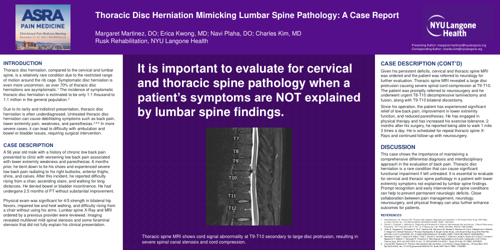
Given his persistent deficits, cervical and thoracic spine MRI was ordered and the patient was referred to neurology for further evaluation. Thoracic spine MRI revealed a large disc protrusion causing severe spinal cord compression at T9-T10. The patient was promptly referred to neurosurgery and he underwent urgent T8-T10 decompressive laminectomy and fusion, along with T9-T10 bilateral discectomy.
Since his operation, the patient has experienced significant relief of low back pain, improvement in lower extremity function, and reduced paresthesias. He has engaged in physical therapy and has increased his exercise tolerance. 2 months after his surgery, he reported being able to walk 1 mile 3 times a day. He is scheduled for repeat thoracic spine X-Rays and continued follow-up with neurosurgery.
DISCUSSION
This case shows the importance of maintaining a comprehensive differential diagnosis and interdisciplinary approach in the evaluation of back pain. Thoracic disc herniation is a rare condition that can cause significant functional impairment if left untreated. It is essential to evaluate for cervical and thoracic spine pathology in a patient with lower extremity symptoms not explained by lumbar spine findings. Prompt recognition and early intervention of spine conditions can help to prevent permanent neurologic deficits. Close collaboration between pain management, neurology, neurosurgery, and physical therapy can also further enhance outcomes for patients.