501 posters, 2 videos, 1 audios, 8 topics, 37 sessions, 395 authors, 307 institutions
ePostersLive by SciGen Technologies S.A. All rights reserved.
23rd Annual Pain Medicine Meeting 2024
November 21-23, 2024 | Caesars Palace, Las Vegas, Nevada

5820
INADVERTENT ACCESS OF THE RETRODURAL SPACE OF OKADA DURING A FLUOROSCOPIC GUIDED LUMBAR TRANSFORAMINAL EPIDURAL STEROID INJECTION
Poster Presenter
Authors
Affiliations
Part of Topic
Medically Challenging Cases
Inadvertent Access of the Retrodural Space of Okada During a Fluoroscopic Guided Lumbar Transforaminal Epidural Steroid Injection
Introduction:
The retrodural space of Okada is a potential space posterior to the ligamentum flavum that allows communication with the bilateral facet joints. However, the actual anatomy of this space has not been clearly visualized to date (1).
Methods:
Outpatient and inpatient procedure notes were reviewed.
As the case report is devoid of patient identifiable information, it is exempt from IRB review requirements as per Jefferson Moss-Magee Rehabilitation Hospital policy.
Case Report:
We present the case of a 77-year-old female with past surgical history of posterior lumbar interbody fusion (PLIF) at L4-5 in 2016 who initially presented to the outpatient office with years of progressively worsening chronic low back pain with radicular pain along the anterolateral aspect of her entire right lower extremity along with worsening right lower extremity weakness. Initial physical exam was pertinent for right hip flexor weakness, graded 4/5 along with decreased sensation to light touch along the right lower extremity L2 and L3 dermatomes. Magnetic resonance imaging (MRI) of the lumbar spine without contrast was ordered which was most notable for a right paracentral disc extrusion with caudal migration at the L1-2 level, effacing the right lateral recess and displacing the cauda equina nerve roots posteriorly as well as diffuse facet arthrosis. Patient deferred surgical evaluation and elected to proceed with a planned right L2-3 transforaminal epidural steroid injection (TFESI).
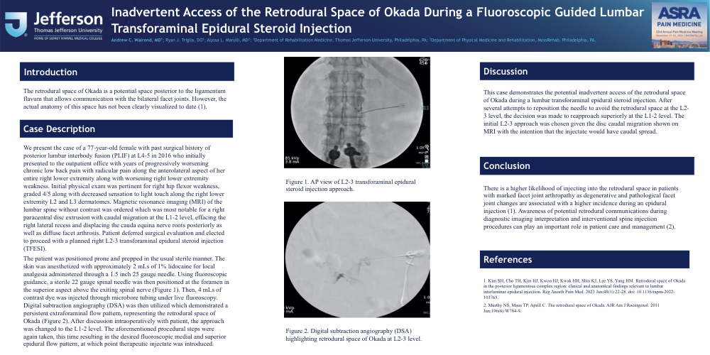
The patient was positioned prone and prepped in the usual sterile manner. The skin was anesthetized with approximately 2 mLs of 1% lidocaine for local analgesia administered through a 1.5 inch 25 gauge needle. Using fluoroscopic guidance, a sterile 22 gauge spinal needle was then positioned at the foramen in the superior aspect above the exiting spinal nerve (Image 1). Then, 4 mLs of contrast dye was injected through microbore tubing under live fluoroscopy. Digital subtraction angiography (DSA) was then utilized which demonstrated a persistent extraforaminal flow pattern, representing the retrodural space of Okada (Image 2). After discussion intraoperatively with patient, the approach was changed to the L1-2 level. The aforementioned procedural steps were again taken, this time resulting in the desired fluoroscopic medial and superior epidural flow pattern, at which point therapeutic injectate was introduced.
Discussion:
This case demonstrates the potential inadvertent access of the retrodural space of Okada during a lumbar transforaminal epidural steroid injection. After several attempts to reposition the needle to avoid the retrodural space at the L2-3 level, the decision was made to reapproach superiorly at the L1-2 level. The initial L2-3 approach was chosen given the disc caudal migration shown on MRI with the intention that the injectate would have caudal spread.
There is a higher likelihood of injecting into the retrodural space in patients with marked facet joint arthropathy as degenerative and pathological facet joint changes are associated with a higher incidence during an epidural injection (1). Awareness of potential retrodural communications during diagnostic imaging interpretation and interventional spine injection procedures can play an important role in patient care and management (2).