501 posters, 2 videos, 1 audios, 8 topics, 37 sessions, 395 authors, 307 institutions
ePostersLive by SciGen Technologies S.A. All rights reserved.
23rd Annual Pain Medicine Meeting 2024
November 21-23, 2024 | Caesars Palace, Las Vegas, Nevada

6301
: IMPLANTATION OF CERVICAL PERIPHERAL NERVE STIMULATOR FOR TREATMENT OF OCCIPITAL NEURALGIA
Poster Presenter
Authors
Affiliations
Part of Topic
Medically Challenging Cases
Title: Implantation of Cervical Peripheral Nerve Stimulator for Treatment of Occipital Neuralgia
Authors: Matthew Potter MD, Yawar J. Qadri, MD PhD
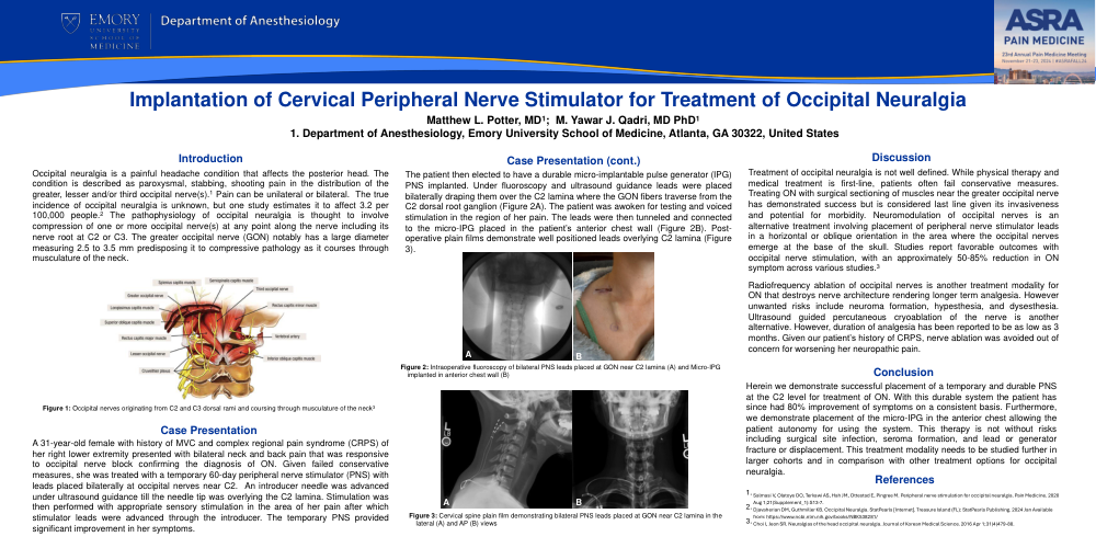
Introduction: Occipital neuralgia is a painful headache condition that affects the posterior head. The condition is described as paroxysmal, stabbing, shooting pain in the distribution of the greater, lesser and/or third occipital nerve(s).1 Pain can be unilateral or bilateral. The true incidence of occipital neuralgia is unknown, but one study estimates it to affect 3.2 per 100,000 people.2 The pathophysiology of occipital neuralgia is thought to involve compression of one or more occipital nerve(s) at any point along the nerve including its nerve root at C2 or C3. The greater occipital nerve (GON) notably has a large diameter measuring 2.5 to 3.5 mm predisposing it to compressive pathology as it courses through musculature of the neck.
Case Presentation: A 31-year-old female with history of MVC and complex regional pain syndrome (CRPS) of her right lower extremity presented with bilateral neck and back pain that was responsive to occipital nerve block confirming the diagnosis of ON. Given failed conservative measures, she was treated with a temporary 60-day peripheral nerve stimulator (PNS) with leads placed bilaterally at occipital nerves near C2. An introducer needle was advanced under ultrasound guidance till the needle tip was overlying the C2 lamina. Stimulation was then performed with appropriate sensory stimulation in the area of pain after which stimulator leads were advanced through the introducer. The temporary PNS provided significant improvement in her symptoms.
The patient then elected to have a durable micro-implantable pulse generator (IPG) PNS implanted. Under fluoroscopy and ultrasound guidance leads were placed bilaterally draping them over the C2 lamina where the GON fibers traverse from the C2 dorsal root ganglion (Figure 2A). The patient was awoken for testing and voiced stimulation in the region of her pain. The leads were then tunneled and connected to the micro-IPG placed in the patient’s anterior chest wall (Figure 2B). Post-operative plain films demonstrate well positioned leads overlying C2 lamina (Figure 3).
Discussion: Treatment of occipital neuralgia is not well defined. While physical therapy and medical treatment is first-line, patients often fail conservative measures. Treating ON with surgical sectioning of muscles near the greater occipital nerve has demonstrated success but is considered last line given its invasiveness and potential for morbidity. Neuromodulation of occipital nerves is an alternative treatment involving placement of peripheral nerve stimulator leads in a horizontal or oblique orientation in the area where the occipital nerves emerge at the base of the skull. Studies report favorable outcomes with occipital nerve stimulation, with an approximately 50-85% reduction in ON symptom across various studies.3
Radiofrequency ablation of occipital nerves is another treatment modality for ON that destroys nerve architecture rendering longer term analgesia. However unwanted risks include neuroma formation, hypesthesia, and dysesthesia. Ultrasound guided percutaneous cryoablation of the nerve is another alternative. However, duration of analgesia has been reported to be as low as 3 months. Given our patient’s history of CRPS, nerve ablation was avoided out of concern for worsening her neuropathic pain.
Conclusion: Herein we demonstrate successful placement of a temporary and durable PNS at the C2 level for treatment of ON. With this durable system the patient has since had 80% improvement of symptoms on a consistent basis. Furthermore, we demonstrate placement of the micro-IPG in the anterior chest allowing the patient autonomy for using the system. This therapy is not without risks including surgical site infection, seroma formation, and lead or generator fracture or displacement. This treatment modality needs to be studied further in larger cohorts and in comparison with other treatment options for occipital neuralgia.
References:
- Salmasi V, Olatoye OO, Terkawi AS, Hah JM, Ottestad E, Pingree M. Peripheral nerve stimulation for occipital neuralgia. Pain Medicine. 2020 Aug 1;21(Supplement_1):S13-7.
- Djavaherian DM, Guthmiller KB. Occipital Neuralgia. StatPearls [Internet]. Treasure Island (FL): StatPearls Publishing. 2024 Jan Available from: https://www.ncbi.nlm.nih.gov/books/NBK538281/
- Choi I, Jeon SR. Neuralgias of the head occipital neuralgia. Journal of Korean Medical Science. 2016 Apr 1;31(4)479-88.
Figure 1: Occipital nerves originating from C2 and C3 dorsal rami and coursing through musculature of the neck
Figure 2: Intraoperative fluoroscopy of bilateral PNS leads placed at GON near C2 lamina (A) and Micro-IPG implanted in anterior chest wall (B)
Figure 3: Cervical spine plain film demonstrating bilateral PNS leads placed at GON near C2 lamina in the lateral (A) and AP (B) views