501 posters, 2 videos, 1 audios, 8 topics, 37 sessions, 395 authors, 307 institutions
ePostersLive by SciGen Technologies S.A. All rights reserved.
23rd Annual Pain Medicine Meeting 2024
November 21-23, 2024 | Caesars Palace, Las Vegas, Nevada

6092
A RARE COMPLICATION: A CASE OF LARGE SUBCUTANEOUS HEMATOMA POST SCS IMPLANTATION MIGRATING FROM POCKET TO PARAMEDIAN INCISION
Poster Presenter
Authors
Affiliations
Part of Topic
Medically Challenging Cases
Introduction
Chronic low back pain (CLBP) is a global health issue leading to substantial disability despite numerous therapeutic interventions [1]. Spinal cord stimulation (SCS) is a prominent non-opioid treatment modality, efficacious for conditions refractory to conventional therapies[2]. However, SCS is not devoid of complications: hardware-related, biological, and technical issues. Most common complications: lead migration and device malfunction. Less frequent complications: seroma, epidural abscess and hematoma [3-4]
Methods
Study Type: Case report.
Consent: Signed consent form obtained. IRB Status: Waived.
Results/ Case Report
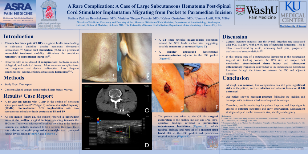
A 65-year-old female with CLBP in the setting of persistent spinal pain syndrome (PSPS type 2) underwent a high-frequency (10kHz) thoracolumbar SCS implantation with two percutaneous stimulator leads contacts at T8 and T9. At one-month follow-up, the patient reported a protruding mass at the midline surgical incision extending towards the IPG site. There was evidence of localized swelling at the lumbar incision site, initially suspected to be a seroma. However, there was substantial rapid progression overnight that prompted further investigation (Figure A and Figure B). A CT scan revealed mixed-density collection around the SCS leads anchor site, suggesting possible hematoma or seroma (Figure C). A doppler ultrasound demonstrated neovascularization adjacent to the IPG pocket (Figure D). The patient was taken to the OR for surgical exploration of the midline incision and IPG. Intra- operative findings revealed a paramedian subcutaneous hematoma (Figure E), which required drainage and removal of a medium-sized blood clot at the IPG pocket and paramedian surgical incision (Figure F).
Discussion
Current literature suggests that the overall infection rate associated with SCS is 2.45%, with a 0.3% rate of neuraxial hematoma. This is often characterized by acute, worsening back pain, progressive weakness and sensory deficits [5]. In this unique case of a subcutaneous hematoma at the paramedian surgical site tracking towards the IPG site, we suspect that mechanical stress-induced tissue injury and subsequent inflammatory responses contributed to the subcutaneous hematoma formation through the interaction between the IPG and adjacent tissues.
Conclusion
Although less common, this complication can still pose significant risks to the patient, such as infection and abscess formation if left untreated. Our patient showed excellent progress following the incision and drainage, with no issues noted in subsequent follow-ups. Therefore, careful monitoring for yellow flags and red flags signs is critical to optimize outcomes and early intervention. Management strategies depend on the hematoma size, stability and urgency.