501 posters, 2 videos, 1 audios, 8 topics, 37 sessions, 395 authors, 307 institutions
ePostersLive by SciGen Technologies S.A. All rights reserved.
23rd Annual Pain Medicine Meeting 2024
November 21-23, 2024 | Caesars Palace, Las Vegas, Nevada

6231
A NOVEL TECHNIQUE FOR RADIOFREQUENCY ABLATION IN CHALLENGING LUMBAR ANATOMY CASES USING CONTINUOUS STIMULATION: A CASE REPORT
Poster Presenter
Authors
Affiliations
NYU Langone Health, New York University Grossman School of Medicine
Part of Topic
Medically Challenging Cases
A Novel Technique for Radiofrequency Ablation in Challenging Lumbar Anatomy Cases Using Continuous Stimulation: A Case Report
Introduction
Radiofrequency ablation (RFA) of the lumbar medial branch nerves is a widely used treatment for facetogenic lower back pain, as endorsed by the American Society of Pain and Neuroscience (ASPN) 2022 consensus guidelines.1 However, this procedure becomes increasingly challenging in cases of complex lumbar anatomy when traditional fluoroscopic anatomy is distorted. This report describes the novel use of continuous low-level stimulation to optimize RFA probe placement based on live patient feedback.
Case Report
A 68-year-old female with a history of scoliosis and osteoporosis presented with chronic lower back pain and right leg radicular pain in an L5/S1 pattern. Her examination revealed right-sided lumbar tenderness, bilateral axial pain with ipsilateral facet loading, and a positive right-sided straight leg raise. The patient reported significant limitations in daily activities, such as difficulty getting into and out of her car due to the lower back pain. Despite extensive physical therapy, pharmacological treatments, and epidural steroid injections, she experienced incomplete relief and continued on ibuprofen 800 mg TID and gabapentin 200 mg nightly.
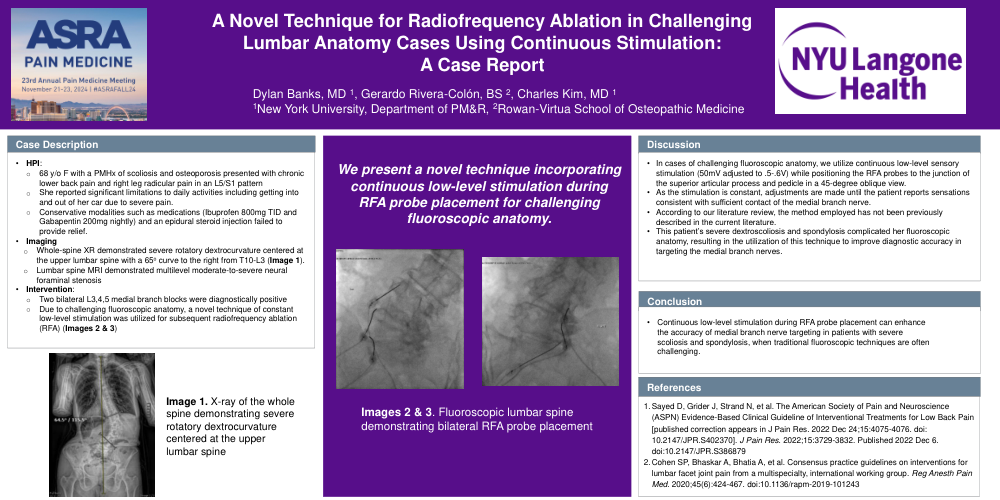
Whole-spine x-rays demonstrated severe rotatory dextrocurvature centered at the upper lumbar spine, with a 65-degree curve to the right from T10 to L3 (Image 1). Lumbar spine MRI showed multilevel moderate-to-severe neural foraminal stenosis. Due to her osteoporosis, surgical interventions were not pursued, and the patient remained on bone growth-promoting medications.
Following two positive diagnostic bilateral lumbar 3, 4, 5 medial branch blocks, RFA was pursued (Images 2, 3). Her severe scoliotic lumbar spine made the transverse process-superior articular process junction difficult to identify on fluoroscopy. Therefore, we used a novel technique involving continuous low-level stimulation while making micro-adjustments to the ablation probes. We utilized sensory stimulation turned to approximately .5 to .6v while the probe was adjusted until the patient reported the sought after sensation consistent with contact along the medial branch nerves. This was used in conjunction with fluoroscopy for placement confirmation.
Discussion
The use of continuous low-level stimulation during RFA probe adjustments is a novel technique we employ when fluoroscopic anatomy is challenging. According to our review, this method has not been previously described in the literature. In this case, the patient’s severe scoliosis and spondylosis complicated her fluoroscopic anatomy, prompting us to use this technique to improve accuracy in targeting her medial branch nerves. The ASPN 2022 consensus guidelines support the use of fluoroscopy for optimizing probe placement, which is considered standard care. Our case introduces a novel approach for cases where typical anatomy is difficult to identify, integrating current standard of care guidelines with a technique that enhances the accuracy of medial branch nerve identification.1,2