425 posters, 1 videos, 9 audios, 8 topics, 53 sessions, 340 authors, 286 institutions
ePostersLive by SciGen Technologies S.A. All rights reserved.
22nd Annual Pain Medicine Meeting 2023
November 10 - 11, 2023 | New Orleans Marriott | New Orleans, Louisiana, USA

4612
TECHNICAL MODIFICATION OF CERVICAL FACET JOINT RADIOFREQUENCY ABLATION - A NOVEL APPROACH
Poster Presenter
Authors
Affiliations
Part of Topic
Safety/QA/QI Projects
Technical Modification of Cervical Facet Joint Radiofrequency Ablation- A Novel Approach
Introduction
Neck pain is one of the leading causes of disability globally, ranking 21st out of 291 diseases for the total burden of health based on the disability-adjusted life years scale. It has been well established that the cervical facet joints are innervated by the medial branch (MB) of the dorsal ramus spinal nerve. In facet joints below C2-C3, innervation come from articular branches of two medial branches from the dorsal rami at the level of the joint and above the joint.
Currently, dual comparative medial branch blocks (MBBs) are considered by many as the accepted standard for diagnosing cervical facet joint pain. Patients who meet this criteria are candidates for thermal radiofrequency ablation (RFA) of the MB.
Under the guidance of fluoroscopy, both lateral and anterior-posterior (AP) views are used during needle placement to confirm that the electrode is positioned parallel to the targeted MB nerve at the articular pillar of the cervical facet joint. Once placement of the electrode is confirmed on all views, thermal RFA is performed to denervate the facet joint. However, the current cervical thermal RFA technique is oftentimes limited due to shoulder musculature restricting the visibility of the lower cervical facet joints. The technical recommendation described in this paper introduces a modification to the conventional cervical thermal RFA procedure. Specifically, a modified oblique fluoroscopic imaging technique is utilized to increase lower cervical spine facet joint visibility and consequently reduce intraoperative complications and increase patient safety.
Methods
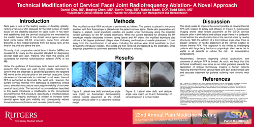
The modified cervical RFA technique is performed as follows: The patient is placed in the prone position. A C-Arm fluoroscope is placed over the patient cervical spine. Sterile skin preparation and draping is applied. Local anesthetic needles are guided under fluoroscopy along the projected needle pathways for the RF needle electrodes. While the current standard for directing the RF introducer needle electrodes involves taking lateral and AP views, the modified technique also utilizes a 50 degree ipsilateral oblique view. Following confirmation of needle placement 1-2 mm away from the neuroforamen in all views, including the oblique view, local anesthetic is given through the introducer needles. The stylets are then removed and replaced by the electrodes. Once electrode placement is confirmed, standard RFA protocol is followed.
Results
Figure 1. Lateral view (left) and oblique angle view (right) on fluoroscopy demonstrating optimal needle placement at the C5-C6 cervical articular pillar in a cadaveric skeletal model.
Figure 2. Lateral view (left) and oblique angle view (right) on C-arm fluoroscopy of cervical spine in a human model.
Discussion
This study seeks to improve the current practice of cervical thermal RFA with respect to safety and efficacy. In Figure 1, fluoroscopic imaging shows ideal needle placement at the C5-C6 cervical articular pillar in both lateral and oblique angle views in a cadaveric model without the visual obstruction of the cervical spine by nearby structures. With the addition of a third oblique angle view, there is greater certainty of needle placement at the correct location to initiate thermal RFA. This approach is not limited to challenging patients with large body habitus or physiologic short necks but is viable in all patients to reduce the risk of intraoperative complications.
Nevertheless, literature regarding clinical applications and outcomes of oblique RFA is limited. As such, we hope that this technical modification can serve as an initial guideline towards the application of oblique fluoroscopy imaging in human patients receiving thermal RFA for cervical facet joint pain and promote safe and accurate treatment for patients suffering from chronic neck pain.