425 posters, 1 videos, 9 audios, 8 topics, 53 sessions, 340 authors, 286 institutions
ePostersLive by SciGen Technologies S.A. All rights reserved.
22nd Annual Pain Medicine Meeting 2023
November 10 - 11, 2023 | New Orleans Marriott | New Orleans, Louisiana, USA

4826
MRI RF HEATING OF SCS LEADS: ENSURING PATIENT SAFETY THROUGH LEAD DESIGN, SIMULATION AND ANALYSIS
Poster Presenter
Authors
Affiliations
Part of Topic
Emerging Technology
MRI RF Heating of SCS Leads: Ensuring Patient Safety Through Lead Design, Simulation and Analysis
Magnetic Resonance Imaging (MRI) is an important diagnostic imaging technique and is the recommended diagnostic modality for many clinical indications. Up to 98% of spinal cord stimulation (SCS) patients may need an MRI scan within 10 years of implant.1 However, creating MR Conditional labeling for SCS devices that ensures patient safety and maximizes clinical diagnostic utility is challenging because of the complexity of the MRI environment. Radiofrequency (RF) lead heating can potentially result in serious patient harm but is particularly difficult to evaluate because it is a function of the lead design, patient attributes, and scan conditions.
This poster describes the RF heating modeling and test methods used to support full-body Normal Operating Mode MR Conditional labeling for a novel RF shielded SCS lead designed to reduce RF electrode heating. The methods are compliant with Clause 8, Tier 3, of ISO/TS 10974:20182 that contains industry consensus test methods for evaluating implanted device safety in the MRI environment.
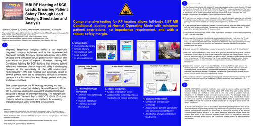
The risk of patient injury due to MRI related RF heating is evaluated using a transfer function (TF) lead model method (Clause 8, Tier 3, of ISO/TS 10974:2018). The TF is a measured property of the lead that determines its propensity to heat when exposed to RF energy. Once calibrated and validated, the TF lead model can be used to predict lead electrode temperatures for any simulated MRI scan scenario. The approach is illustrated in Figure 1 and summarized below:
TF lead electrode heating models are measured with a specially designed apparatus and calibrated in vitro by measuring the lead model response to known RF exposures. The calibration error and part-to-part lead model variability due to manufacturing tolerance is determined.
The acceptance criterion is a probability of histologically detectable thermal tissue damage for 30-minutes of continuous scanning at the Normal Operation Mode SAR limits of less than 1 in 1 million.
ISO/TS 10974:2018 compliant methods were used to assess safety scanning RF shielded SCS at the Normal Operating Mode SAR limits. The lead heating transfer function models are verified in vitro and in vivo and used to predict electrode temperatures in 180,857 human simulated scan scenarios. The simulated scans are conservative with respect to clinical scan protocols because they consist of 30-minutes of continuous scanning at the Normal Operating Mode limits as opposed to typical clinical MRI protocols that consist of multiple short scans at varying RF power levels with delays in-between.
A Monte Carlo analysis is used to account for lead heating model prediction error and to demonstrate that the probability of inducing detectable tissue thermal damage in human patients conservatively exposed to 30-minutes of continuous scanning at the Normal Operating Mode limits is less than 1 per million. Moreover, additional analysis shows that broken lead wires in the tantalum shielded leads result in insignificant electrode temperature increase and supports MR Conditional labeling without a lead impedance requirement
References
1Desai MJ, Hargens LM, Breitenfeldt MD, Doth AH, Ryan MP, Gunnarsson C, Safriel Y. The rate of magnetic resonance imaging in patients with spinal cord stimulation. Spine (Phila Pa 1976). 2015 May 1;40(9):E531-7. D
2ISO/TS. 2018. ISO/TS 10974: assessment of the safety of magnetic resonance imaging for patients with an active implantable medical device.
3https://www.itis.ethz.ch/virtual-population/virtual-population/overview/ (Accessed Aug. 8,2023)