127 posters, 2 topics, 24 sessions, 326 authors, 85 institutions
ePostersLive by SciGen Technologies S.A. All rights reserved.
2023 ACP ANNUAL SESSION
October 25-28 | San Diego, CA

84
Influence of arch form on the accuracy of 3-D printed dental casts: an in vitro study.
Poster Presenter
Authors
Affiliations
Part of Topic
Original Research
Effect of Varying Arch Forms on the Accuracy of 3-D Printed Dental Casts: An In-Vitro Study
The field of dentistry has witnessed a remarkable transformation in recent years, with the advent of cutting-edge technologies such as 3-D printing. This transformative technology has found applications in various aspects of dentistry, including the fabrication of dental casts, which play a pivotal role in the diagnosis, treatment planning, and prosthodontic restoration of patients.1 Although additively manufactured casts have shown excellent accuracy, the influence of the dental arch form on the accuracy of digital and printed casts has not been studied.2 Considering that different ethnic groups and populations display variable dental arch measurements and characteristics, it is important to assess if the accuracy of digital and 3-D printed casts can be impacted by different arch forms.3,4 According to Chuck’s classification presented in 1934, the dental arch can present three different forms: ovoid, tapered, and square shape.4
This research aims to investigate the accuracy of 3-D printed dental casts from digital data, focusing on the impact of different arch forms (square, tapered, and ovoid). The cost-effectiveness and precision of additive manufacturing offer promising potential in dentistry, but current literature lacks substantial investigation on how arch form variations affect cast accuracy.
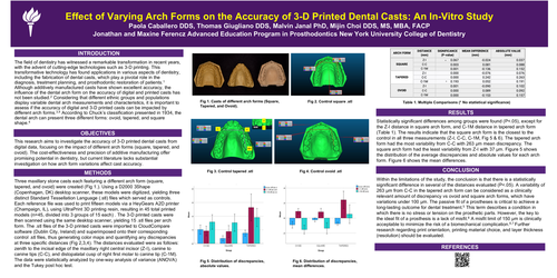
Three maxillary stone casts each featuring a different arch form (square, tapered, and ovoid) were created (Fig 1.). Using a D2000 3Shape (Copenhagen, DK) desktop scanner, these models were digitized, yielding three distinct Standard Tessellation Language (.stl) files which served as controls. Each reference file was used to print fifteen models via a HeyGearsA2D printer (Champaign, IL), using UltraPrint 3D printing resin, resulting in 45 total printed models (n=45, divided into 3 groups of 15 each) . The 3-D printed casts were then scanned using the same desktop scanner, yielding 15 .stl files per arch form. The .stl files of the 3-D printed casts were imported to CloudCompare software (Dublin City, Ireland) and superimposed onto their corresponding control .stl files, thus generating color maps and quantifying any discrepancies at three specific distances (Fig 2,3,4). The distances evaluated were as follows: zenith to the incisal edge of the maxillary right central incisor (Z-I), canine to canine tips (C-C), and distopalatal cusp of right first molar to canine tip (C-1M). The data were statistically analyzed by one-way analysis of variance (ANOVA) and the Tukey post hoc test.
Statistically significant differences among groups were found (P<.05), except for the Z-I distance in square arch form, and C-1M distance in tapered arch form (Table 1). The results indicate that the square arch form is the closest to the control in all three measurements (Z-I, C-C, C-1M, Fig 5 & 6). The tapered arch form had the most variability from C-C with 263 μm mean discrepancy. The square arch form had the least variability from Z-I with 37 μm. Figure 5 shows the distribution of the average discrepancies and absolute values for each arch form. Figure 6 shows the mean differences.
Within the limitations of the study, the conclusion is that there is a statistically significant difference in several of the distances evaluated (P<.05). A variability of 263 μm from C-C in the tapered arch form can be considered as a clinically relevant amount of discrepancy vs ovoid and square arch forms, which have variations under 100 μm. The passive fit of a prostheses is critical to achieve a long-lasting outcome for dental treatment.5 This term describes a condition in which there is no stress or tension on the prosthetic parts. However, the key to the ideal fit of a prosthesis is a lack of misfit.6 A misfit limit of 150 μm is clinically acceptable to minimize the risk of a biomechanical complication.6,7 Further research regarding print orientation, printing material choice, and layer thickness (resolution) should be evaluated.