127 posters, 2 topics, 24 sessions, 326 authors, 85 institutions
ePostersLive by SciGen Technologies S.A. All rights reserved.
2023 ACP ANNUAL SESSION
October 25-28 | San Diego, CA

196
Literature Review: Evolution of Dental Adhesives- Past, Present and Future.
Poster Presenter
Authors
Affiliations
Part of Topic
Original Research
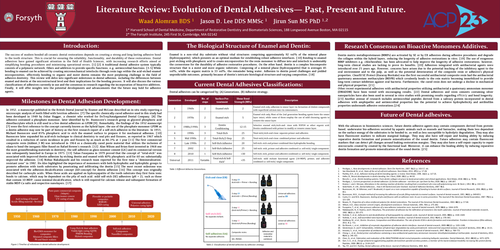
Literature Review: Evolution of Dental Adhesives— Past, Present and Future.
introduction:
The success of modern bonded all-ceramic dental restorations depends on creating a strong and long-lasting adhesive bond
to the tooth structure. This is crucial for ensuring the reliability, functionality, and durability of these restorations. Dental
adhesives have gained significant attention in the field of Health Sciences, with increasing research efforts aimed at
simplifying bonding procedures and minimizing operational errors. [1] [2] A traditional dental adhesive system typically
consists of a polymeric network, fillers and additives to provide specific functions, e.g. antimicrobial functions. [3-5] While
bonding to enamel can be achieved by creating micromechanical interlocks through resin tags within the acid-etched enamel
microporosities, effectively bonding to organic and moist dentin remains the most perplexing challenge in the field of
adhesive dentistry. This review will delve into significant milestones in dental adhesion, including the differences between
enamel and dentin at the microstructural level and their implications for the bonding process. It will also discuss the various
classifications of adhesives currently in use and the consensus in research regarding the incorporation of bioactive additives.
Finally, it will offer insights into the potential developments and advancements that the future may hold for adhesive agents.
Milestones in Dental Adhesion Development:
In 1952, a manuscript published in the British Dental Journal by Kramer and McLean described an in-situ study reporting a
microscopic analysis of teeth filled with Sevriton adhesive. [7] The specific chemically-cured adhesive used in this study had
been developed in 1949 by Oskar Hagger, a chemist who worked for DeTrey/Amalgamated Dental Company. [8] The
adhesive contained a phosphate monomer, later identified by Dr. Buonocore’s research group as glycerol phosphoric acid
dimethacrylate which is still used in a few dental adhesives as GPDM [9]. Remarkably, the findings of the 1952 manuscript
[7] were the first reference to the concept currently known as the hybrid layer, the use of the phosphate monomer GPDM as
a dentin adhesive may now be part of history as the first research report of a self-etch adhesive in the literature. In 1955,
Michael Buonocore used 85% phosphoric acid to etch the enamel surface to prepare it for mechanical adhesion. [10]
Buonocore later expanded his acid-etch technique to clinical dentistry to seal pits and fissures, as reported in 1967 which
resulted in a reduction of caries incidence in pits and fissures by as much as 86.3% at 1 year [11] The first commercial
composite resin (Addent,3 M) was introduced in 1964 as a chemically cured paste material that utilizes the inclusion of
silane to bond the inorganic filler Based on Rafael Bowen’s research. [12] Alan Wilson and Brian Kent invented in 1968 one
the most groundbreaking materials in dentistry, the self-adhesive as glass-ionomer cement (GIC) and its commercial version
was subsequently launched in Europe in 1975. [13] Takao Fusayama in 1979 proved that the concept of “total-etch” was
associated with improved dentin adhesion by etching dentin and enamel with 40% phosphoric acid for 60 sec substantially
improved the adhesion. [14] Nobuo Nakabayashi and his research team reported for the first time a “demineralizationresistant
zone” in 1982. He Also highlighted the importance of monomers with both hydrophobic and hydrophilic groups to
promote adhesion with tooth substrates by penetrating and infiltrating the dentin [15] The most recent milestone was
associated with the adhesion-decalcification concept (AD-concept) for dentin adhesion [16] This concept was originally
described for carboxylic acids. When these acids are applied on hydroxyapatite of the tooth substrate they first form ionic
bonds to calcium, which may be dependent on the pKa of each acid. mild self-etch (SE) adhesives (pH ≈ 2), such as those
that contain 10-MDP, cause minimal decalcification, which is still required required for calcium release and subsequent formation of
stable MDP-Ca salts and respective nanolayers. [17]
The Biological Structure of Enamel and Dentin:
Enamel is a non-vital dry substrate without vital structures comprising approximately 92 vol% of the mineral phase
(hydroxyapatite), which presents as an optimal medium for establishing robust adhesive interfaces. [10] Bonding to enamel
post etching with phosphoric acid to create microporosities for the resin monomer to diffuse into and interlock is undeniably
the cornerstone for the durability of adhesive restorative procedures. On the other hand, dentin is a complex biocomposite
structure that is a moist and more organic substrate. Composing of a mineral phase (hydroxyapatite) that is averages 45
vol%, while the organic matrix is 33 vol%, the remainder being water. Adhesion to dentin posed challenges and yielded
unpredictable outcomes, primarily because of dentin's intricate histological structure and varying composition. [18]
Research Consensus on Bioactive Monomers Additives.
Dentin matrix metalloproteinases (MMPs) are activated by SE or by ER adhesives during adhesive procedures and degrade
the collagen within the hybrid layers, reducing the longevity of adhesive restorations in vitro. [19] The use of exogenous
MMP inhibitors e.g. chlorhexidine, has been advocated to help improve the longevity of adhesive restorations, however,
long-term clinical studies are lacking to prove its benefits. [20] Adhesives integrated with antibacterial agents were
introduced over 25 years ago, However, concerns have arisen due to the potential leaching of the inhibitory agent to the
oral environment and to the pulp, as well as the potential deterioration of the composite resin or adhesive's physical
properties. Clearfil SE Protect (Kuraray Noritake) was the first successful antibacterial composite resin had the antibacterial
quaternary ammonium methacrylate (MDPB) which covalently bonds to the rein matrix becoming immobilized to provide
long-term contact-inhibition against oral bacteria. Furthermore, the cured resin does not undergo a decline in mechanical
and physical properties. [21]
Other recent experimental adhesives with antibacterial properties utilizing antibacterial a quaternary ammonium monomer
(DMADDM) have been tested with encouraging results. [22] Dental adhesives and resin cements containing silver
nanoparticles have also been used in several in vitro studies with promising results regarding efficacy and bond longevity.
[23] Another avenue of research includes antimicrobial peptides derived from a salivary protein incorporated in dental
adhesives with amphipathic and antimicrobial properties has the potential to achieve hydrophobicity and antibiofilm
properties underneath adhesive restorations.[24]
Future of Dental adhesives.
With the advances in biomimetics sciences, future dentin adhesive agents may contain components derived from protein-based,
underwater bio-adhesives secreted by aquatic animals such as mussels and barnacles, making them less dependent
on the surface energy of the substrates to be bonded to, as well as less susceptible to hydrolytic degradation. They may also
have fluorescent markers to indicate marginal leakage. They may also have self-repair and healing ability by inducing
reparative dentin formation and promote mineralization of the sound dentin. Future adhesives may contain fluorescent
markers that can detect pH changes around leaking restoration margins. They may also have a self-repair capacity to repair
microcracks created by created by the functional load. Moreover, it can enhance the healing ability by inducing reparative
dentin formation and promote mineralization of the sound dentin. [25]