105 posters, 2 topics, 24 sessions, 264 authors, 71 institutions
ePostersLive by SciGen Technologies S.A. All rights reserved.
2022 ACP Annual Session
2 - 5 November | Austin, TX

76
Occlusion Changes upon Crown Cementation with Different Cement Space Settings
Poster Presenter
Authors
Affiliations
DUMMY, Harvard School of Dental Medicine, Harvard School of Dental Medicine
Part of Topic
Original Research
Occlusion Changes upon Crown Cementation with Different Cement Space Settings
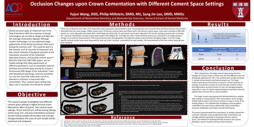
introduction: Dental occlusion plays an important role in the field of dentistry. With the evolution of dental technologies, we are able to design and fabricate full coverage restorations digitally. Although modern technology is at very advanced stage, adjustments of the dental prostheses often occur during the insertion visit1. This could be due to a few reasons, such as accuracy of impression and bite record, intensity of designed occlusion of the restoration, precision of the restoration fabrication process, and possibly cement space2,3. With the help from CAD-CAM system, we can modify settings that allow adjustments of different parameters, such as restorative material thickness and cement space during the virtual 3-dimensional (3D) design of the restoration3. Even with detailed pre-plannings, clinicians sometime run into the issue that crown does not fit as planned or occlusion is inaccurate after cementation. Thus, cement space setting may play a role in occlusion and should be studied.
Objective:
This research project investigates how different cement space settings in digital Zirconia crown fabrications affect occlusion. Two cement space settings, 70 μm and 120 μm, will be testing settings of this study. Pre- and post-cementation occlusion of each testing sample will be taken and scanned. Changes between the scans of each sample will be examined and compared.
Methods:
Thirty sets of identical casts with a first molar (#3) crown preparation were printed in resin. The preparation was scanned (Trios) and thirty Zirconia crowns were fabricated from the same design. Fifteen crowns had a 70 microns cement space and fifteen had a 120 microns cement space. Casts were mounted in MIP with plaster on a semi adjustable articulator with a fixed hinge axis (Artex CR). Pre-cemented crowns were adjusted to fit into the existing occlusion prior to testing. Pre- and post-cementation occlusal records were made in PVS (Regisil 2X). A DC light box with a fixed overhead camera was connected to an image analyzer (Image J) and used for measurements. PVS occlusal records were photographed in the light box station and converted into digital images. From the image, specific occlusal contact areas were measured for each set of pre- and post- cementation for each sample. The measurements of the two cement space groups were analyzed.
Conclusion:
After comparisons, the larger cement space group had less changes of occlusal contact surface area, but the difference was not statistically significant (P<.05 ). This could be due to large variations in sample measurements and limited sample size. The larger cement space group also had less increase in contact intensity on average. Increasing in contact area and intensity could be critical and might induce occlusal trauma if it was not managed properly, especially in cases of cement retained implant restorations because implant does not have PDL as shock absorber4.
The larger cement space group also demonstrated less samples with change in occlusal pattern after cementation. Some changes shifted contact points from fossa to inclines of cusps in some of the testing subjects. This indicated that changing in occlusal pattern could affect the cusp to fossa relationship, which could affect occlusion stabilization and induce occlusal interference.
The project demonstrated that cementation of samples altered the occlusion. Clinically, occlusal adjustment after cementation was strongly advised, especially for the group with less cement space. The method of analyzing PVS bite records in this project provided a way to visualize and document occlusion.