105 posters, 2 topics, 24 sessions, 264 authors, 71 institutions
ePostersLive by SciGen Technologies S.A. All rights reserved.
2022 ACP Annual Session
2 - 5 November | Austin, TX

63
CATER; A DIGITAL SINGLE TOOTH IMPLANT STUDY
Poster Presenter
Authors
Mariana Fernandez Lozada, Sandra Al-Tarawneh, Ghadeer Thalji, Cooper Lyndon
Affiliations
Part of Topic
Original Research
CATER; A DIGITAL SINGLE TOOTH IMPLANT STUDY
Fernandez Lozada M1, Al-Tarawneh S 1 , Thalji G1, Cooper LF2
1 Department of Restorative Dentistry, College of Dentistry, UIC, Chicago, IL, USA2 Department of Oral Biology College of Medicine, UIC, Chicago, IL, USA
INTRODUCTION
Implant success requires both sufficient bone alveolar volume and sufficient mucosal tissue to protect against biofilm-mediated inflammation, assure functional comfort and contribute to dental implant esthetics.1 Following dental implant placement, the alveolar ridge resorbs approximately 0.5 to 1.0 mm during the first year. Healed ridges are also often volumetrically deficient, losing up to 30% of their horizontal volume following tooth extraction.2
Peri-implant mucosal grafting has been proposed and is currently part of dental implant therapy.3,4 Both autogenous grafts (mucosal connective tissue from the roof of the patient's mouth or "palate")5 and allogeneic grafts (collagen-rich grafts derived from a genetically non-identical human donor) are used for these purposes, but there is controversy regarding the effectiveness of supplemental graft use during implant treatment. 6
HYPOTHESIS/OBJECTIVE
Comprehensive Alveolar Therapy and Esthetic Rehabilitation (CATER) for single-tooth implant requires compensation for alveolar resorption. We hypothesize that an allogenic soft-tissue augmentation enhances peri-implant tissue contour and esthetics.
METHODS
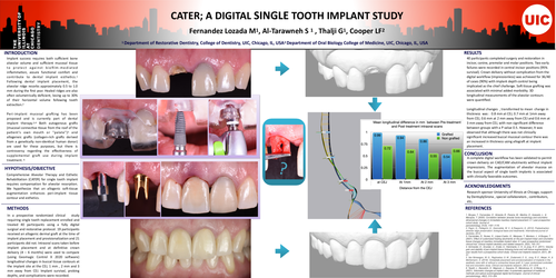
In a prospective randomized clinical study requiring single tooth replacement, enrolled and treated 40 participants using a fully digital surgical and restorative protocol. 19 participants received an allogenic dermal graft at the time of implant placement and provisionalization, and 21 participants did not. Intraoral scans taken before implant placement and at definitive crown delivery (4 – 6 months) were used to compare (using Geomagic Control X 2020 software) longitudinal changes in buccal tissue contours at the implant site at the CEJ, 1 mm, 2 mm, and 3 mm away from CEJ. Implant survival, probing depths, and complications were recorded.
RESULTS
40 participants completed surgery and restoration in incisor, canine, premolar, and molar positions. Two early failures were recorded in central incisor positions (95% survival). Crown delivery without complication from the digital workflow (impressionless) was achieved for 36/40 of cases (90%), with implant depth control being implicated as the chief challenge. Soft tissue grafting was associated with minimal added morbidity. 3D longitudinal measurements of the alveolar contours were quantified.
Longitudinal changes, transformed to mean change in thickness, was: 0.8 mm at CEJ, 0.7 mm at 1mm away from CEJ, 0.6 mm at 2 mm away from CEJ, and 0.6 mm at 3 mm away from CEJ, with a nonsignificant difference between groups with a P value 0.5. However, It was observed that although there was no clinically significant increase in buccal mucosal contour, there was an increase in thickness using allograft at implant placement.
Conclusion
A complete digital workflow has been validated to permit crown delivery on CAD/CAM abutments without implant impressions. The augmentation of alveolar mucosa on the buccal aspect of single-tooth implants is associated with clinically favorable outcomes.
Acknowledgments
The research sponsor is the University of Illinois at Chicago, supported by Dentsply Sirona, special collaborators, contributors, etc.
REFERENCES
1. Borges, T., Fernandes, D., Almeida, B., Pereira, M., Martins, D., Azevedo, L., & Marques, T. (2020). Correlation between alveolar bone morphology and volumetric dimensional changes in immediate maxillary implant placement: A 1-year prospective cohort study. Journal of
periodontology, 91(9), 1167–1176.
2. Pagni, G., Pellegrini, G., Giannobile, W. V., & Rasperini, G. (2012). Postextraction alveolar ridge preservation: biological basis and treatments. International journal of dentistry, 2012,
3. Fernandes, D., Nunes, S., López-Castro, G., Marques, T., Montero, J., & Borges, T. (2021). Effect of customized healing abutments on the peri-implant linear and volumetric tissue changes at maxillary immediate implant sites: A 1-year prospective randomized clinical trial. Clinical implant dentistry and related research, 23(5), 745–757.
4. Schneider, D., Grunder, U., Ender, A., Hämmerle, C. H., & Jung, R. E. (2011). Volume gain and stability of peri-implant tissue following bone and soft tissue augmentation: 1-year results from a prospective cohort study. Clinical oral implants research, 22(1), 28–37.
5. Van Nimwegen, W. G., Raghoebar, G. M., Zuiderveld, E. G., Jung, R. E., Meijer, H., & Mühlemann, S. (2018). Immediate placement and provisionalization of implants in the aesthetic zone with or without a connective tissue graft: A 1-year randomized controlled trial and volumetric study. Clinical oral implants research, 29(7), 671–678.
6. Tavelli, L., Barootchi, S., Majzoub, J., Siqueira, R., Mendonça, G., & Wang, H. L. (2021). Volumetric changes at implant sites: A systematic appraisal of traditional methods and optical scanningbased digital technologies. Journal of clinical periodontology, 48(2), 315–334.