105 posters, 2 topics, 24 sessions, 264 authors, 71 institutions
ePostersLive by SciGen Technologies S.A. All rights reserved.
2022 ACP Annual Session
2 - 5 November | Austin, TX

143
<p>Influence of Gingival Retraction Cord Position on the Accuracy of Intraoral</p> <p>Scanner for Subgingival Finish Line</p>
Poster Presenter
Authors
Affiliations
Part of Topic
Original Research
Influence of Gingival Retraction Cord on the Accuracy of Intraoral Scanner for Subgingival Finish Line
Objectives
A previous clinical pilot study showed that intraoral scanners have limited predictability in scanning subgingival finish lines(1). Therefore, supragingival finish lines or the use of gingival retraction were recommended to improve accuracy of digital impressions(2).
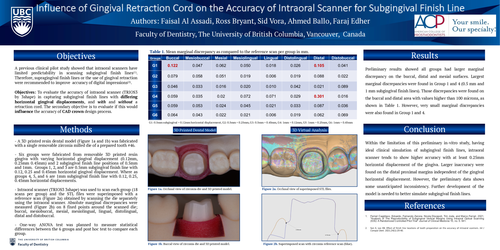
Objectives: To evaluate the accuracy of intraoral scanner (TRIOS3 by 3shape) in capturing subgingival finish lines with differing horizontal gingival displacements, and with and without a retraction cord. The secondary objective is to evaluate if this would influence the accuracy of CAD crown design process.
Methods
- A 3D printed resin dental model (Figure 1a and 1b) was fabricated with a single removable zirconia milled die of a prepared tooth #46.
- Six groups were fabricated from removable 3D printed resin gingiva with varying horizontal gingival displacement (0.12mm, 0.25mm 0.45mm) and 2 subgingival finish line positions of 0.5mm and 1mm. Groups 1, 2, and 3 are 0.5mm subgingival finish line with 0.12, 0.25 and 0.45mm horizontal gingival displacement. Where as groups 4, 5, and 6 are 1mm subgingival finish line with 0.12, 0.25, 0.45mm horizontal displacements.
- Intraoral scanner (TRIOS3 3shape) was used to scan each group (18 scans per group) and the STL files were superimposed with a reference scan (Figure 2a) obtained by scanning the die separately using the intraoral scanner. Absolute marginal discrepancies were measured (Figure 2b) on 8 fixed points around the scanned die; buccal, mesiobuccal, mesial, mesiolingual, lingual, distolingual, distal and distobuccal.
- One-way ANOVA test was planned to measure statistical differences between the 6 groups and post hoc test to compare each group.
Preliminary results showed all groups had larger marginal discrepancy on the buccal, distal and mesial surfaces. Largest marginal discrepancies were found in Group 1 and 4 (0.5 mm and 1 mm subgingival finish lines). Those discrepancies were found on the buccal and distal area with values higher than 100 microns, as shown in Table 1. However, very small marginal discrepancies were also found in Group 1 and 4.
Conclusion
Within the limitation of this preliminary in-vitro study, having ideal clinical simulation of subgingival finish lines, intraoral scanner tends to show higher accuracy with at least 0.25mm horizontal displacement of the gingiva. Larger inaccuracy were found on the distal proximal margins independent of the gingival horizontal displacement. However, the preliminary data shows some unanticipated inconsistency. Further development of the model is needed to better simulate subgingival finish lines.