241 posters, 10 topics, 7 sessions, 899 authors, 296 institutions
ePostersLive by SciGen Technologies S.A. All rights reserved.
The AAPS 103rd Annual Meeting 2025
May 24-27, 2025 | Austin, Texas

P7
The Angiogenic Effect Of Micropuncture On Arterial And Venous Endothelial Cells
Part of Topic
Basic Science Research
The Angiogenic Effect of Micropuncture on Arterial and Venous Endothelial Cells
Mohammad Hossein Asgardoon, MD, MPH, Shailaja Agrawal, PhD, Mary Landmesser, BS , Maryam Abdelaal, BS, Jazzmyn Dawes, BS, Dino Ravnic, DO, MPH
Pennsylvania State University, College of Medicine, Hershey, PA, USA
Introduction
•Scaffolds are cornerstone to reconstructive surgery and tissue engineering.
•Their success is limited by slow vascularization following implantation.
•Micropuncture (MP) is a novel surgical approach that can be used to induce angiogenesis and expedite adjacent scaffold vascularization.
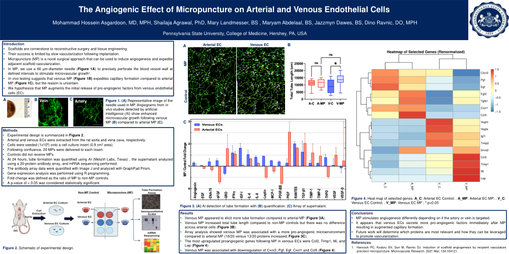
•In MP, we use a 60 µm-diameter needle (Figure 1A) to precisely perforate the blood vessel wall at defined intervals to stimulate microvascular growth1.
•In vivo testing suggests that venous MP (Figure 1B) expedites capillary formation compared to arterial MP (Figure 1C), but the reason is uncertain.
•We hypothesize that MP augments the initial release of pro-angiogenic factors from venous endothelial cells (EC).
Methods
•Experimental design is summarized in Figure 2.
•Arterial and venous ECs were extracted from the rat aorta and vena cava, respectively.
•Cells were seeded (1x10⁵) onto a cell culture insert (0.9 cm2 area).
•Following confluence, 20 MPs were delivered to each insert.
•Controls did not receive MPs.
•At 24 hours, tube formation was quantified using AI (MetaVi Labs, Texas) , the supernatant analyzed using a 20-protein antibody array, and mRNA sequencing performed.
•The antibody array data were quantified with Image J and analyzed with GraphPad Prism.
•Gene expression analysis was performed using R programming.
•Fold change was defined as the ratio of MP to non-MP controls.
•A p-value of < 0.05 was considered statistically significant.
Results
•Venous MP appeared to elicit more tube formation compared to arterial MP (Figure 3A).
•Venous MP increased total tube length compared to non-MP controls but there was no difference across arterial cells (Figure 3B).
•Array analysis showed venous MP was associated with a more pro-angiogenic microenvironment compared to arterial MP (18/20 versus 13/20 proteins increased; Figure 3C).
•The most upregulated proangiogenic genes following MP in venous ECs were Ccl2, Timp1, Il6, and Lep (Figure 4).
•Venous MP was associated with downregulation of Cxcr2, Pgf, Egf, Cxcl1 and Ccl5 (Figure 4).
Conclusions
•MP stimulates angiogenesis differently depending on if the artery or vein is targeted.
•It appears that venous ECs secrete more pro-angiogenic factors immediately after MP resulting in augmented capillary formation.
•Future work will determine which proteins are most relevant and how they can be leveraged to promote vascularization.