241 posters, 10 topics, 7 sessions, 899 authors, 296 institutions
ePostersLive by SciGen Technologies S.A. All rights reserved.
The AAPS 103rd Annual Meeting 2025
May 24-27, 2025 | Austin, Texas

D14
Comparing Non-invasive Perfusion Measuring Modalities In Predicting Skin Injury Following Flap Creation
Part of Topic
Basic Science Research
Title: Comparing Non-Invasive Perfusion Measuring Modalities in Predicting Skin Injury Following Flap Creation
Authors: David A. Febre-Alemañy1,2,6,Shreeniket Pawar3,4,Bijay Ghimirey3,4,Ji Ho Park1,2,Neekita Jikaria1,2 ,Anilchandra Attaluri3,4,Mohammad-Reza Tofighi3,4 ,Dino Ravnic1,2,5
Affiliations:
1Irvin S. Zubar Plastic Surgery Research Laboratory, Penn State College of Medicine, Hershey, Pennsylvania, USA.
2Department of Surgery, Penn State Health Milton S. Hershey Medical Center, Hershey, Pennsylvania, USA.
3Department of Mechanical Engineering, School of Science, Engineering, and Technology, The Pennsylvania State University Harrisburg, Harrisburg , Pennsylvania, USA.
4Department of Electrical Engineering, School of Science, Engineering, and Technology, The Pennsylvania State University Harrisburg, Harrisburg Pennsylvania, USA.
5Huck Institutes of the Life Sciences, The Pennsylvania State University, University Park, State College, Pennsylvania, USA.
6San Juan Bautista School of Medicine, Caguas, Puerto Rico, USA.
Purpose:
Flaps are crucial in reconstructive surgery, but inadequate perfusion can lead to necrosis and compromise outcomes. Multiple non-invasive modalities have been developed for assessing perfusion intraoperatively during flap elevation, but the optimal modality to predict necrosis progression remains unclear.
Methods:
Six random pattern skin-flaps were created on the dorsum of four domestic pigs, with each flap having a contralateral non-flap control. Both sides were divided into six smaller zones for analysis, with zone 1 being the most proximal and zone 6 being most distal. Oxygen saturation, temperature, and total hemoglobin index (THI) were measured in all zones immediately following flap creation. At Day 7, animals were euthanized and skin thickness was quantified in each zone using hematoxylin and eosin staining. Statistical analysis was performed using unpaired t-tests (α = 0.05).
Results:
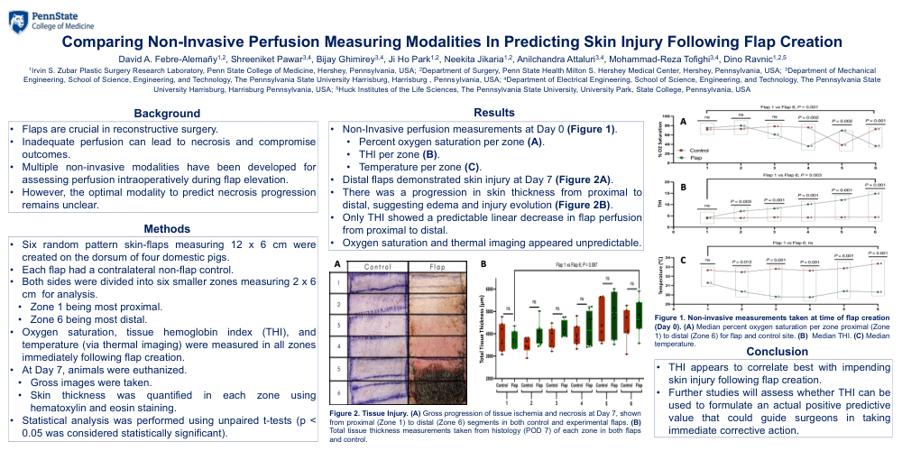
Only THI showed a predictable linear decrease in flap perfusion from proximal to distal compared to controls at Day 0. Oxygen saturation and thermal imaging appeared unpredictable. At Day 7, there was a progression in skin thickness from proximal to distal, suggesting edema and injury evolution. Results are summarized in Figure 1.
Conclusion:
THI appears to correlate best with impending skin injury following flap creation. Further studies are needed to assess whether THI can be used to formulate an actual positive predictive value that could guide surgeons in taking immediate corrective action.