241 posters, 10 topics, 7 sessions, 899 authors, 296 institutions
ePostersLive by SciGen Technologies S.A. All rights reserved.
The AAPS 103rd Annual Meeting 2025
May 24-27, 2025 | Austin, Texas

D16
Evaluating The Safety Of Transdermal Deferoxamine Patch In Oncological Settings
Poster Presenter
Authors
Affiliations
Part of Topic
Basic Science Research
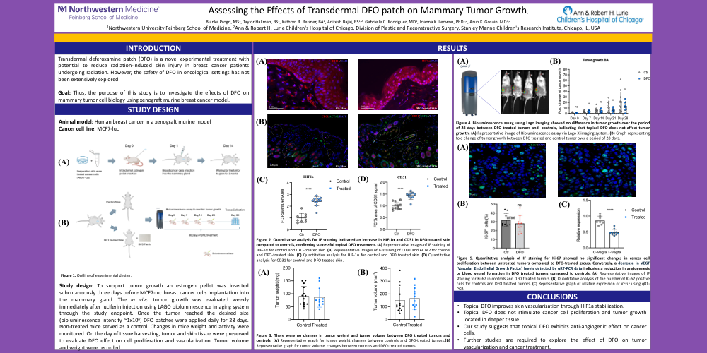
Transdermal deferoxamine patch (DFO) is a novel experimental treatment with potential to reduce radiation-induced skin injury in breast cancer patients undergoing radiation. However, the safety of DFO in oncological settings has not been extensively explored.
Goal: Thus, the purpose of this study is to investigate the effects of DFO on mammary tumor cell biology using xenograft murine breast cancer model.
Animal model: Human breast cancer in a xenograft murine model
Cancer cell line: MCF7-luc
Figure 1. Outline of experimental design
Study design: To support tumor growth an estrogen pellet was inserted subcutaneously three days before MCF7-luc breast cancer cells implantation into the mammary gland. The in vivo tumor growth was evaluated weekly immediately after luciferin injection using LAGO bioluminescence imaging system through the study endpoint. Once the tumor reached the desired size (bioluminescence intensity ~1x10⁸) DFO patches were applied daily for 28 days. Non-treated mice served as a control. Changes in mice weight and activity were monitored. On the day of tissue harvesting, tumor and skin tissue were preserved to evaluate DFO effect on cell proliferation and vascularization. Tumor volume and weight were recorded.
Figure 2. Quantitative analysis for IF staining indicated an increase in HIF-1α and CD31 in DFO-treated skin compared to controls, confirming successful topical DFO treatment. (A) Representative images of IF staining of HIF-1α for control and DFO-treated skin. (B) Representative images of IF staining of CD31 and ACTA2 for control and DFO-treated skin. (C) Quantitative analysis for HIF-1α for control and DFO treated skin. (D) Quantitative analysis for CD31 for control and DFO treated skin.
Figure 3. There were no changes in tumor weight and tumor volume between DFO treated tumors and controls. (A) Representative graph for tumor weight changes between controls and DFO-treated tumors.(B) Representative graph for tumor volume changes between controls and DFO-treated tumors.
Figure 4. Bioluminescence assay, using Lago imaging showed no difference in tumor growth over the period of 28 days between DFO-treated tumors and controls, indicating that topical DFO does not affect tumor growth. (A) Representative image of Bioluminescence assay via Lago X imaging system. (B) Graph representing fold change of tumor growth between DFO treated and control tumor over a period of 28 days.
Figure 5. Quantitative analysis of IF staining for Ki-67 showed no significant changes in cancer cell proliferation between untreated tumors compared to DFO-treated group. Conversely, a decrease in VEGF (Vascular Endothelial Growth Factor) levels detected by qRT-PCR data indicates a reduction in angiogenesis or blood vessel formation in DFO treated tumors compared to controls. (A) Representative images of IF staining for Ki-67 in controls and DFO treated tumors. (B) Quantitative analysis of the number of Ki-67 positive cells for controls and DFO treated tumors. (C) Representative graph of relative expression of VEGF using qRT-PCR.
Conclusions: